

Suppression of cathepsins B and L causes a proliferation of lysosomes and the formation of meganeurites in hippocampus
- PMID: 9151717
- PMCID: PMC6573562
- DOI: 10.1523/JNEUROSCI.17-11-04006.1997
Suppression of cathepsins B and L causes a proliferation of lysosomes and the formation of meganeurites in hippocampus
Abstract
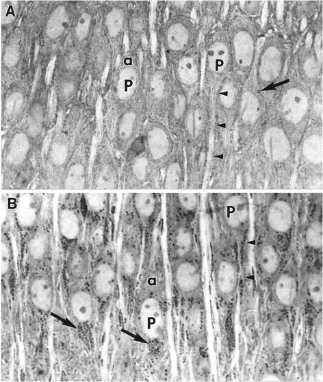
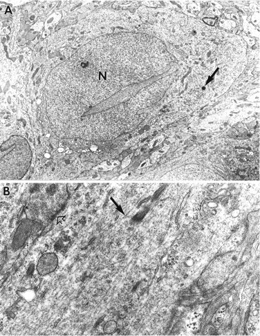
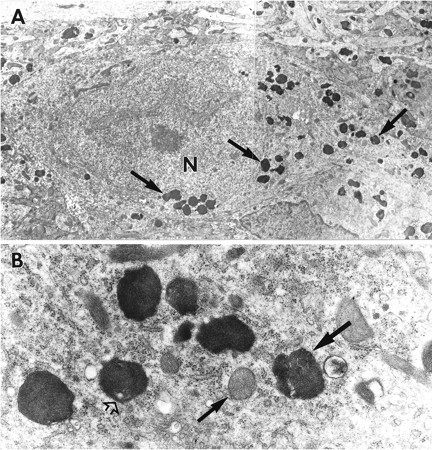
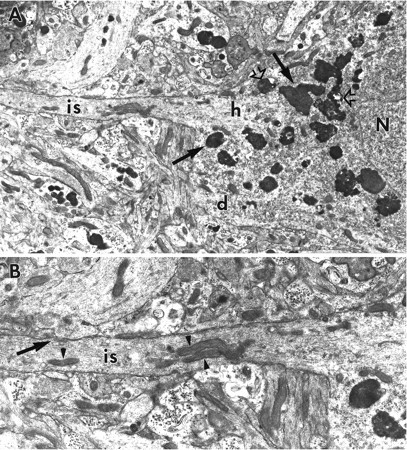
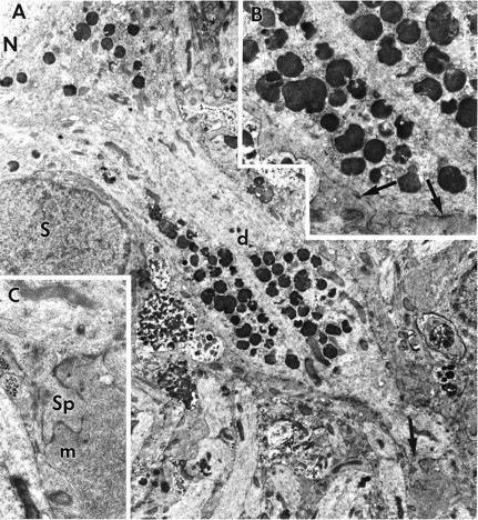
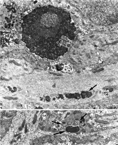
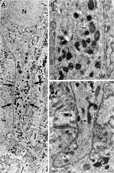
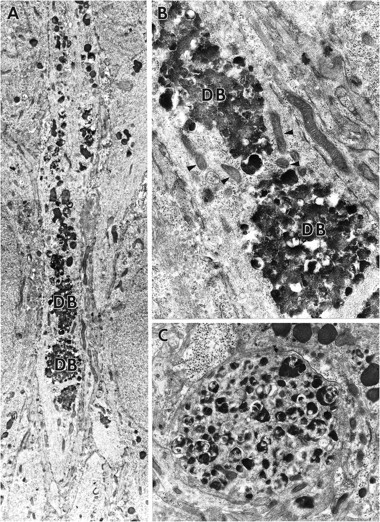
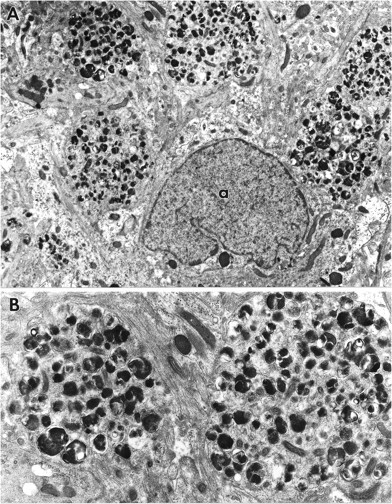
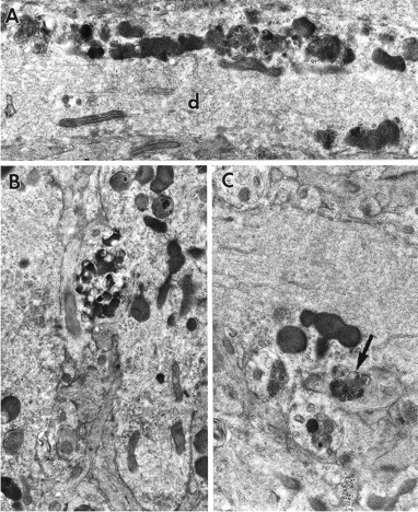
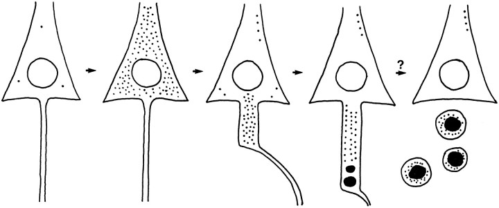
Cultured hippocampal slices exhibited prominent ultrastructural features of brain aging after exposure to an inhibitor of cathepsins B and L. Six days of treatment with N-CBZ-L-phenylalanyl-L-alanine-diazomethylketone (ZPAD) resulted in a dramatic increase in the number of lysosomes in the perikarya of neurons and glial cells throughout the slices. Furthermore, lysosomes in CA1 and CA3 pyramidal cells were not restricted to the soma but instead were located throughout dendritic processes. Clusters of lysosomes were commonly found within bulging segments of proximal dendrites that were notable for an absence of microtubules and neurofilaments. Although pyknotic nuclei were sometimes encountered, most of the cells in slices exposed to ZPAD for 6 d appeared relatively normal. Slices given 7 d of recovery contained several unique features, compared with those processed immediately after incubation with the inhibitor. Cell bodies of CA1 neurons were largely cleared of the excess lysosomes but had gained fusiform, somatic extensions that were filled with fused lysosomes and related complex, dense bodies. These appendages, similar in form and content to structures previously referred to as "meganeurites," were not observed in CA3 neurons or granule cells. Because meganeurites were often interposed between cell body and axon, they have the potential to interfere with processes requiring axonal transport. It is suggested that inactivation of cathepsins B and L results in a proliferation of lysosomes and that meganeurite generation provides a means of storing residual catabolic organelles. The accumulated material could be eliminated by pinching off the meganeurite but, at least in some cases, this action would result in axotomy. Reduced cathepsin L activity, increased numbers of lysosomes, and the formation of meganeurites are all reported to occur during brain aging; thus, it is possible that the infusion of ZPAD into cultured slices sets in motion a greatly accelerated gerontological sequence.
Figures
References
-
- Bahr BA, Abai B, Gall C, Vanderklish PW, Hoffman KB, Lynch G. Induction of β-amyloid-containing polypeptides in hippocampus: evidence for a concomitant loss of synaptic proteins and interactions with an excitotoxin. Exp Neurol. 1995a;129:81–94. - PubMed
-
- Bahr BA, Kessler M, Rivera S, Vanderklish PW, Hall RA, Mutneja MS, Gall C, Hoffman KB. Stable maintenance of glutamate receptors and other synaptic components in long-term hippocampal slices. Hippocampus. 1995b;5:425–439. - PubMed
-
- Bahr BA, Tiriveedhi S, Park GY, Lynch G. Induction of calpain-mediated spectrin fragments by pathogenic treatments in long-term hippocampal slices. J Pharmacol Exp Ther. 1995c;273:902–908. - PubMed
-
- Barrett AJ, Kirschke H. Cathepsin B, cathepsin H, and cathepsin L. Methods Enzymol. 1981;80:535–561. - PubMed
-
- Bednarski E, Lynch G. Cytosolic proteolysis of tau by cathepsin D in hippocampus following suppression of cathepsins B and L. J Neurochem. 1996;67:1846–1855. - PubMed
Publication types
MeSH terms
Substances
Grants and funding
LinkOut - more resources
Full Text Sources
Other Literature Sources
Miscellaneous