. 1997 Jun 1;17(11):4212-22.
doi: 10.1523/JNEUROSCI.17-11-04212.1997.

Alzheimer's presenilin mutation sensitizes neural cells to apoptosis induced by trophic factor withdrawal and amyloid beta-peptide: involvement of calcium and oxyradicals

Affiliations

Alzheimer's presenilin mutation sensitizes neural cells to apoptosis induced by trophic factor withdrawal and amyloid beta-peptide: involvement of calcium and oxyradicals

Q Guo et al. J Neurosci. .

Abstract

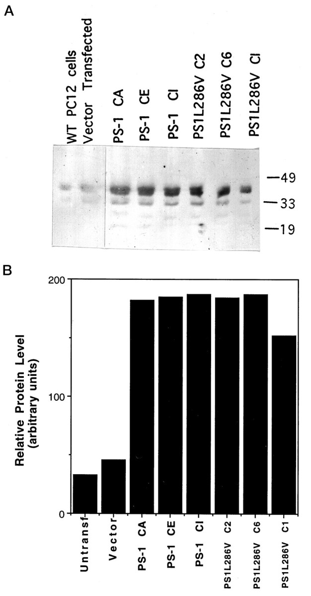
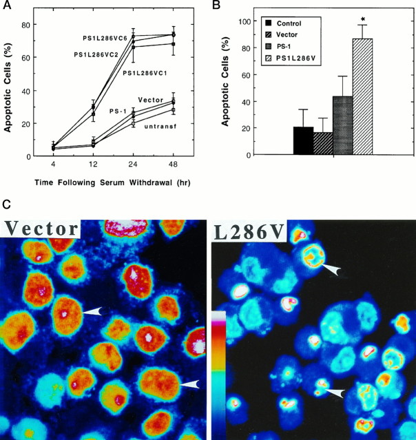
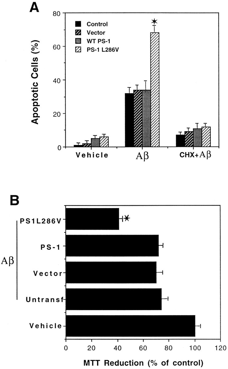
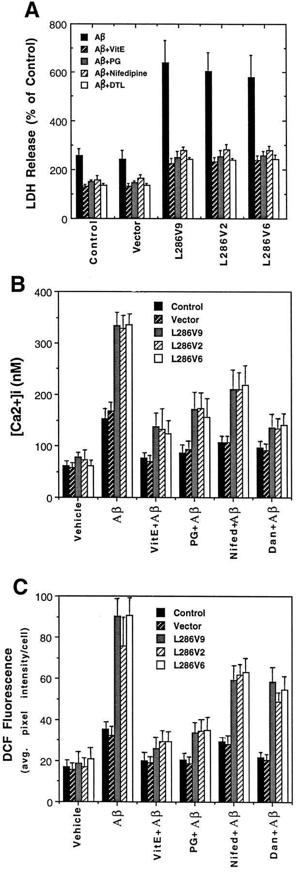
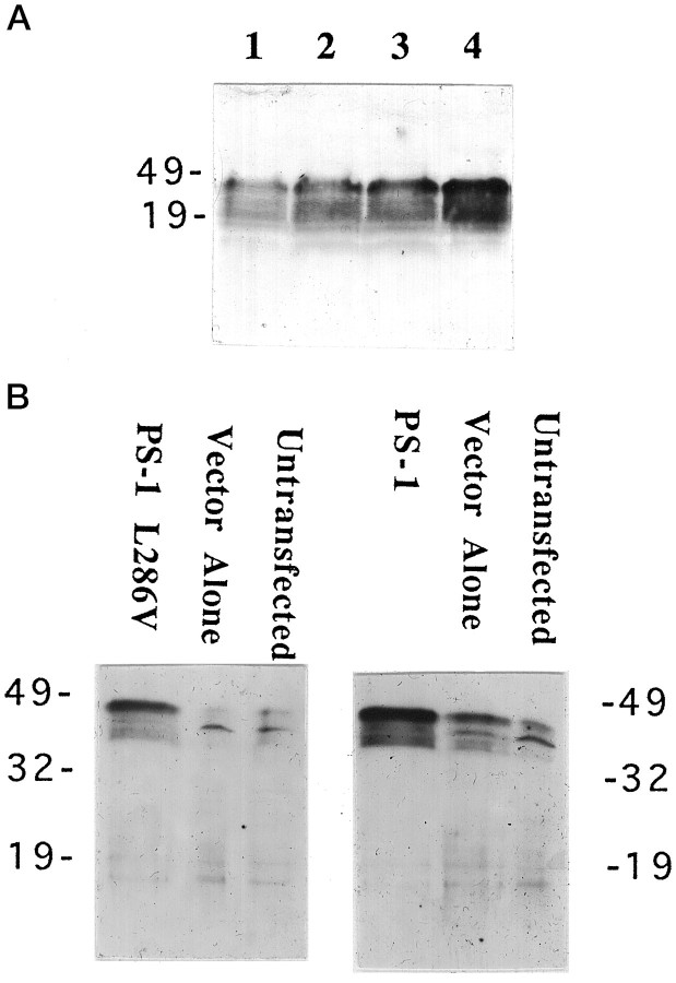
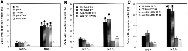
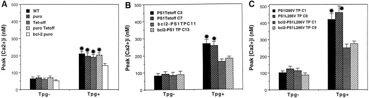
Most autosomal dominant inherited forms of early onset Alzheimer's disease (AD) are caused by mutations in the presenilin-1 (PS-1) gene on chromosome 14. PS-1 is an integral membrane protein with six to nine membrane-spanning domains and is expressed in neurons throughout the brain wherein it is localized mainly in endoplasmic reticulum (ER). The mechanism or mechanisms whereby PS-1 mutations promote neuron degeneration in AD are unknown. Recent findings suggest links among deposition of amyloid beta-peptide (Abeta), oxidative stress, disruption of ion homeostasis, and an apoptotic form of neuron death in AD. We now report that expression of the human PS-1 L286V mutation in PC12 cells increases their susceptibility to apoptosis induced by trophic factor withdrawal and Abeta. Increases in oxidative stress and intracellular calcium levels induced by the apoptotic stimuli were exacerbated greatly in cells expressing the PS-1 mutation, as compared with control cell lines and lines overexpressing wild-type PS-1. The antiapoptotic gene product Bcl-2 prevented apoptosis after NGF withdrawal from differentiated PC12 cells expressing mutant PS-1. Elevations of [Ca2+]i in response to thapsigargin, an inhibitor of the ER Ca2+-ATPase, were increased in cells expressing mutant PS-1, and this adverse effect was abolished in cells expressing Bcl-2. Antioxidants and blockers of calcium influx and release from ER protected cells against the adverse consequences of the PS-1 mutation. By perturbing cellular calcium regulation and promoting oxidative stress, PS-1 mutations may sensitize neurons to apoptotic death in AD.


Figures

Fig. 1.
Fig. 1.
Expression of wild-type and mutant PS-1 in PC12 cells. A, Western blot showing PS-1 protein in untransfected and vector-transfected PC12 cells and in three different lines of cells expressing wild-type (PS-1) and mutant (PS1L286V) PS-1. Equivalent amounts of protein (50 μg/lane) from cell homogenates of the indicated cell lines (untransfected cells, vector-transfected cells, PS-1, and PS1L286V) were separated by SDS-PAGE, transferred to nitrocellulose, and probed with PS-1 antibody. Similar to results of other investigators (Thinakaran et al., 1996), in addition to recognizing full-length PS-1 (46 kDa band), our anti-loop PS-1 antibody also recognized presumptive proteolytic products of PS-1 ∼32 and 19 kDa. B, Densitometric analysis of relative levels of PS-1 protein in PC12 cell lines stably expressing wild-type (PS-1) and mutant (PS1L286V) PS-1. Note that basal PS-1 levels are relatively low in PC12 cells and that levels of expression of wild-type and mutant PS-1 were similar among the lines shown.
Fig. 2.
Fig. 2.
PC12 cells expressing PS-1 L286V mutation exhibit increased vulnerability to apoptosis induced by trophic factor withdrawal. A, The percentages of cells exhibiting DNA condensation and fragmentation (apoptotic cells) were determined in Hoescht-stained cultures at the indicated times after serum withdrawal from undifferentiated PC12 cell lines: untransf, untransfected cells; Vector, cells transfected with empty vector; PS-1, cells transfected with wild-type PS-1 (pooled data from three different lines; C1, C2, and C6); and three different lines expressing mutant PS-1 (PS1L286V). Values represent the mean and SD of determinations made in four separate cultures (200 cells counted/culture). Values for each of the three cell lines expressing L286V were greater than corresponding values for untransfected cultures and cultures transfected with vector or wild-type PS-1 (12 hr time point, p < 0.05; 24 and 48 hr time points,p < 0.001); ANOVA with Scheffé’spost hoc tests. B, Control untransfected PC12 cells (Control) and PC12 cells transfected with empty vector (Vector), wild-type PS-1 (PS-1), or mutant PS-1 (PS1L286V) were differentiated in the presence of NGF (data pooled from analyses on all three lines expressing wild-type PS-1 and all three lines expressing PS-1 L286V; compare with Fig. 1). NGF was withdrawn, and 48 hr later the percentages of apoptotic cells in each culture were determined. Values represent the mean and SD of determinations made in four separate cultures. *p < 0.01 compared with each of the other values; ANOVA with Scheffé’s post hoc tests. C, Confocal images of propidium iodide fluorescence in vector-transfected and L286V-transfected PC12 cells 24 hr after serum withdrawal. Fluorescence intensity is depicted in pseudocolor according to the color scale bar. Note that DNA is distributed diffusely throughout the nuclei of most vector-transfected cells, whereas DNA is fragmented to varying extents in most cells expressing PS-1 L286V (arrowheads).
Fig. 3.
Fig. 3.
Mutant PS-1 increases PC12 cell vulnerability to apoptosis and mitochondrial dysfunction induced by amyloid β-peptide. A, The indicated cell lines were exposed to vehicle (Vehicle), 50 μm Aβ (), or 10 μm cycloheximide plus 50 μm Aβ (CHX + Aβ) for 24 hr. Then cells were stained with Hoescht dye, and the percentages of cells exhibiting DNA condensation and fragmentation were determined. Values represent the mean and SD of determinations made in four separate cultures (data pooled from analyses on all three lines expressing wild-type PS-1 and all three lines expressing PS-1 L286V; compare with Fig. 1). For all cell lines the values for cells exposed to Aβ were significantly greater than values for vehicle or CHX plus Aβ-treated cell lines (p < 0.01). *p < 0.01 compared with each of the other cell lines exposed to Aβ; ANOVA with Scheffé’s post hoc tests. B, Parallel cultures of untransfected control cells (Untransf), vector-transfected cells (Vector), three lines of cells transfected with wild-type PS-1 (PS-1; pooled data), and three lines of mutant PS-1 cells (PS1L286V; pooled data) were exposed for 4 hr to 50 μm Aβ, and relative levels of MTT reduction (a measure of mitochondrial function) were quantified. Values are the mean and SD of determinations made in four separate cultures and are expressed as a percentage of vehicle-treated control (vector-transfected) cells (data pooled from analyses on all three lines expressing wild-type PS-1 and all three lines expressing PS-1 L286V; compare with Fig. 1). There were no differences in basal levels of MTT reduction among the various control, wild-type PS-1-expressing, and mutant PS-1-expressing lines (data not shown). *p < 0.01 compared with corresponding values for untransfected, vector-transfected, and WT PS-1-transfected lines exposed to Aβ; ANOVA with Scheffé’s post hoctests.
Fig. 4.
Fig. 4.
PC12 cells expressing PS-1 L286V mutation exhibit increased levels of oxidative stress and intracellular calcium after exposure to Aβ: attenuation by antioxidants and blockers of calcium influx and release from intracellular stores. A, Cultures were pretreated for 24 hr with 50 μm vitamin E (VitE) or for 2 hr with 5 μm propyl gallate (PG), 1 μm nifedipine (Nifedipine), or 1 μm dantrolene (DTL). Then cultures were exposed to 50 μm Aβ for 24 hr, and the medium was removed for LDH assay (cells exposed to Aβ first undergo apoptosis, followed by secondary necrosis, the latter being detected by LDH release assay). Values are expressed as a percentage of the maximal LDH release (mean and SD of 6–8 cultures); maximal LDH release was determined in parallel cultures (of each cell line) subjected to freeze–thaw. Values for each of the Aβ-treated cell lines expressing PS-1 L286V were significantly greater than each of the values for control (untransfected) and vector-transfected cell lines (p < 0.01) and for each of the values in PS-1 L286V cultures pretreated with vitamin E, propyl gallate, nifedipine, or dantrolene (p < 0.01 in each case); ANOVA with Scheffé’s post hoc tests.B, C, Cultures were pretreated with antioxidants or Ca2+ flux blockers as described forA and then exposed to vehicle or Aβ for 4 hr. Then the relative [Ca2+]i (fura-2 imaging) (B) and levels of peroxides (DCF Fluorescence) (C) in individual cells were quantified. Values are the mean and SD of determinations made in three to four cultures (15–25 cells for [Ca2+]imeasurements and 40–60 cells/culture for DCF measurements). Values for each of the Aβ-treated cell lines expressing L286V were significantly greater than each of the values in the vehicle-treated cultures (p < 0.01), each of the values in the cultures pretreated with vitamin E or propyl gallate (p < 0.01), and each of the values for L286V cells in the cultures pretreated with nifedipine or dantrolene (p < 0.05); ANOVA with Scheffé’spost hoc tests.
Fig. 5.
Fig. 5.
Controlled expression of PS-1 in PC12 cells with the use of a tetracyline-responsive transactivator. A, A PC12 cell line expressing the “Tet-off” construct was stably transfected with a pTRE-derived plasmid expressing PS-1 L286V gene. Cells were incubated for 48 hr in the presence of 2.0, 0.004, 0.002, and 0 μg/ml tetracycline (lanes 1–4, respectively). Cell proteins were separated by SDS-PAGE (100 μg/lane), transferred to a nitrocellulose sheet, and immunoreacted with PS-1 antibody. Note that, as the concentration of tetracycline was decreased, the levels of mutant PS-1 expression increased. B, Western blot showing that in the absence of tetracycline a double-stable PS-1 L286V cell line (C1) and a double-stable PS-1 cell line (C3) show significantly higher levels of PS-1 expression than do vector-transfected or untransfected cell lines.
Fig. 6.
Fig. 6.
Mutant PS-1 increases the vulnerability of differentiated PC12 cells to NGF withdrawal-induced apoptosis. Cultures of differentiated PC12 cells were incubated for 48 hr in serum-free medium containing or lacking NGF, and the percentage of cells exhibiting nuclear condensation and fragmentation was quantified. Values are the mean and SEM of determinations made in at least four separate cultures. A, Analyses in various control PC12 cell lines: WT, untransfected wild-type cells;puro, vector-transfected cells (pBabe-puro vector used for Bcl-2 expression); Tet-off, cells transfected with the Tet-off plasmid; puro Tet-off, cells doubly transfected with pBabe-puro vector and Tet-off plasmid; bcl-2 puro, cells expressing Bcl-2. Note that NGF withdrawal induced a similar level of apoptosis in all control lines, whereas cells expressing Bcl-2 were resistant to NGF withdrawal-induced apoptosis. *p < 0.01 compared with corresponding values for NGF+ cultures and the value for Bcl-2 NGFcells. B, Bcl-2 protects PC12 cells overexpressing wild-type PS-1 against NGF withdrawal-induced apoptosis. Two lines of control cells expressing wild-type PS-1 (C3 andC7) and two different lines of cells expressing both Bcl-2 and PS-1 (C11 and C13) were analyzed. *p < 0.01 compared with corresponding values for NGF+ cells and compared with the value for the NGF line expressing Bcl-2. C, Mutant PS-1 enhances vulnerability of PC12 cells to apoptosis induced by NGF withdrawal: protection by Bcl-2. Two lines of control cells expressing PS-1 L286V and two lines of cells expressing both Bcl-2 and PS-1 L286V were analyzed. *p < 0.05 compared with corresponding values for each line expressing wild-type PS-1 (B), p < 0.01 compared with corresponding values for cells maintained in the presence of NGF, andp < 0.01 compared with NGFlines coexpressing Bcl-2; ANOVA with Scheffé’s post hoc tests for pair-wise comparisons.
Fig. 7.
Fig. 7.
Oxidative stress induced by NGF withdrawal is enhanced in PC12 cells expressing mutant PS-1. Cultures of differentiated PC12 cells were incubated for 3 hr in serum-free medium containing or lacking NGF, and levels of cellular peroxides were quantified by confocal laser scanning microscope image analysis of DCF fluorescence. Values are the mean and SEM of determinations made in at least four separate cultures. A, Analyses in various control PC12 cell lines: WT, untransfected wild-type cells; puro, vector-transfected cells;Tet-off, cells transfected with the Tet-off plasmid;puro Tet-off, cells doubly transfected with empty vector and Tet-off plasmid; bcl-2 puro, cells expressing Bcl-2. Note that NGF withdrawal induced a similar level of peroxide accumulation in all control lines and in cells overexpressing Bcl-2. Each value for NGF cultures was significantly greater than the corresponding value for NGF+ cells (p < 0.01). B, Bcl-2 does not prevent NGF withdrawal-induced accumulation of peroxides in PC12 cells overexpressing wild-type PS-1. Two lines of control cells expressing wild-type PS-1 (C3 andC7) and two different lines of cells expressing both Bcl-2 and PS-1 (C11 and C13) were analyzed. Each value for NGF− cultures was significantly greater than the corresponding value for NGF+ cells (p < 0.01). C, Mutant PS-1 enhances accumulation of peroxides in PC12 cells deprived of NGF. Two lines of control cells expressing PS-1 L286V and two lines of cells expressing both Bcl-2 and PS-1 L286V were analyzed. *p < 0.05 compared with corresponding values for each line expressing wild-type PS-1 (B) andp < 0.01 compared with corresponding values for cells maintained in the presence of NGF; ANOVA with Scheffé’spost hoc tests for pair-wise comparisons).
Fig. 8.
Fig. 8.
Elevations of [Ca2+]i induced by thapsigargin are enhanced significantly in PC12 cells expressing mutant PS-1: attenuation by Bcl-2. PC12 cells were incubated in serum-free medium and basal [Ca2+]i (Tpg), and the peak [Ca2+]i after exposure to 1 μm thapsigargin (Tpg+) was quantified (cf. Guo et al., 1996). Values are the mean and SEM of determinations made in at least four separate cultures (15–20 cells/culture). A, Analyses in various control PC12 cell lines: WT, untransfected wild-type cells;puro, vector-transfected cells (vector for Bcl-2-expressing line); Tet-off, cells transfected with the Tet-off plasmid; puro Tet-off, cells doubly transfected with pBabe-puro and Tet-off plasmid; bcl-2 puro, cells expressing Bcl-2. *p < 0.01 compared with corresponding values for Tpg cultures, andp < 0.05 compared with the Tpg+ value in cells expressing Bcl-2. B, The [Ca2+]i response to thapsigargin in cells expressing wild-type PS-1 is attenuated in cells coexpressing Bcl-2. *p < 0.01 compared with corresponding values for Tpg, and p < 0.05 compared with Tpg+ values in cells expressing Bcl-2. C, Mutant PS-1 enhances [Ca2+]i responses to thapsigargin: attenuation by Bcl-2. *p < 0.001 compared with corresponding values for Tpg,p < 0.01 compared with Tpg+ values in cells expressing Bcl-2, and p < 0.01 compared with Tpg+– PS1 Tet-off values (B); ANOVA with Scheffé’s post hoc tests for pair-wise comparisons.

References

    1. Bastitatou A, Greene LA. Aurintricarboxylic acid rescues PC12 cells and sympathetic neurons from cell death caused by nerve growth factor deprivation: correlation with suppression of endonuclease activity. J Cell Biol. 1991;115:461–471. - PMC - PubMed
    1. Behl C, Davis JB, Lesley R, Schubert D. Hydrogen peroxide mediates amyloid β-protein toxicity. Cell. 1994;77:817–827. - PubMed
    1. Benzi G, Moretti A. Are reactive oxygen species involved in Alzheimer’s disease? Neurobiol Aging. 1995;16:661–674. - PubMed
    1. Black MM, Greene LA. Changes in the colchicine susceptibility of microtubules associated with neurite outgrowth: studies with nerve growth factor-responsive PC12 pheochromocytoma cells. J Cell Biol. 1982;95:379–386. - PMC - PubMed
    1. Borchelt DR, Thinakaran G, Eckman CB, Lee MK, Davenport F, Ratovitsky T, Prada C-M, Kim G, Seekins S, Yager D, Slunt HH, Wang R, Seeger M, Levey AI, Gandy SE, Copeland NG, Jenkins NA, Price DL, Younkin SG, Sisodia SS. Familial Alzheimer’s disease-linked presenilin-1 variants elevate Aβ1–42/1–40 ratio in vitro and in vivo. Neuron. 1996;17:1005–1013. - PubMed

Publication types

MeSH terms

LinkOut - more resources