. 1997 Jun 1;17(11):4243-52.
doi: 10.1523/JNEUROSCI.17-11-04243.1997.

Implication of OTX2 in pigment epithelium determination and neural retina differentiation

Affiliations

Implication of OTX2 in pigment epithelium determination and neural retina differentiation

P Bovolenta et al. J Neurosci. .

Abstract

The expression pattern of Otx2, a homeobox-containing gene, was analyzed from the beginning of eye morphogenesis until neural retina differentiation in chick embryos. Early on, Otx2 expression was diffuse throughout the optic vesicles but became restricted to their dorsal part when the vesicles contacted the surface ectoderm. As the optic cup forms, Otx2 was expressed only in the outer layer, which gives rise to the pigment epithelium. This early Otx2 expression pattern was complementary to that of PAX2, which localizes to the ventral half of the developing eye and optic stalk. Otx2 expression was always observed in the pigment epithelium at all stages analyzed but was extended to scattered cells located in the central portion of the neural retina around stage 22. The number of cells expressing Otx2 transcripts increased with time, following a central to peripheral gradient. Bromodeoxyuridine labeling in combination with immunohistochemistry with anti-OTX2 antiserum and different cell-specific markers were used to determine that OTX2-positive cells are postmitotic neuroblasts undergoing differentiation into several, if not all, of the distinct cell types present in the chick retina. These data indicate that Otx2 might have a double role in eye development. First, it might be necessary for the early specification and subsequent functioning of the pigment epithelium. Later, OTX2 expression might be involved in retina neurogenesis, defining a differentiation feature common to the distinct retinal cell classes.


Figures
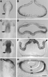
Fig. 1.
Fig. 1.
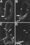
Early expression pattern of Otx2 in the developing chick eye. Chick embryos of stages HH9 (A, B), HH11 (C, D), HH13 (E, F), and HH16 (G, H), subjected to whole-mount in situ hybridization, presented a clear pattern of expression in the anterior portion of the neural tube with a sharp boundary between mesencephalon and rhomboencephalon (arrows in A, C, E, G). Frontal cryostat sections of the stained embryos show the distribution of Otx2 within the eye domain at each developmental stage. Note thatOtx2 is diffuse throughout the optic vesicles at HH9 and HH11 (B, D) but retracts to their dorsal portion at HH13 (arrowhead in F).Otx2 expression in the eye was limited to the prospective pigment epithelium at HH16 (F).lv, Lens vesicle; rn, neural retina; pe, pigment epithelium. Scale bars: A, C, E, 400 μm; G, 500 μm; B, D, F, 40 μm; H, 20 μm.
Fig. 2.
Fig. 2.
Comparison between OTX2 and PAX2 expression pattern in the eye. Confocal microscopic images of frontal cryostat sections through chick eyes at HH18 (A, B) and HH24 (C, D) immunostained with antiserum against OTX2 (A, C) or PAX2 (B, D). Note how pigment and optic stalk epithelial cells are in close proximity (A, B), but a sharp boundary (arrows in A, B) delimits Pax2 and Otx2expression. OTX2 is expressed in the ventral pigment epithelium (C) when PAX2 labels only the optic nerve (D). cb, Ciliary bodies;lv, lens vesicle; le, lens;nr, neural retina; on, optic nerve;os, optic stalk; pe, pigment epithelium;v, ventral. Scale bar, 40 μm.
Fig. 3.
Fig. 3.
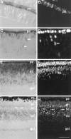
Otx2 mRNA and protein localization in the embryonic chick retina. Frontal cryostat sections of chick retinas hybridized with a digoxigenin-labeled probe (A, C, E) or immunostained with antiserum against OTX2 (B, D, F, H) at E3 (A, B), E3.5 (C, D), E9 (E, F), and E18 (G, H). Otx2 mRNA and protein were detected in a few scattered cells of the central retina (arrows in C, D). The number ofOtx2-positive cells increased with development (E, F). G, Cresyl violet-stained section of a fully differentiated retina in where its layered structure is clearly visible. Note how the nuclei of horizontal cells (arrows in G, H) are OTX2 positive. gcl, Ganglion cell layer; hc,horizontal cells; inl, inner nuclear layer;pe, pigment epithelium; ph,photoreceptors. Scale bars: A–D, 35 μm; E, F, 40 μm; G, H, 60 μm.
Fig. 5.
Fig. 5.
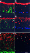
OTX2 is transiently expressed by differentiating RGCs and by cells differentiating into phenotypes other than RGCs. Confocal microscopic images of frontal cryostat sections of chick retinas immunostained with antiserum against OTX2 (red) at E3 (A, B), E5 (C, D), and E7 (E, F) double stained with the RA4 mAb (green in A, C), the anti-islet-1 mAb (blue in B, D), the 3A10 mAb (green in E), or the 3CB2 mAb (green in F). The majority of OTX2-positive cells could be identified as RGCs with the RA4 mAb; only a few cells (arrows) were stained only for OTX2 (A, C). Islet-1-positive RGCs in the mantle do not express OTX2 (C, D). At E7 the majority of OTX2-positive cells are neurons expressing the 3A10 antigen (E). A few OTX2-positive cells can be identified as Müller cells (arrows) by their expression of the 3CB2 antigen (F). fl, Fiber layer;gcl, ganglion cell layer; pe, pigment epithelium. Scale bar, A–D, 30 μm; E, F, 20 μm.
Fig. 4.
Fig. 4.
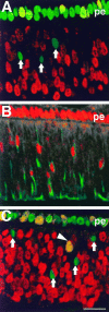
OTX2 is expressed in postmitotic retinal neuroblasts. Confocal microscopic images of frontal cryostat sections of E4 chick retinas fixed 3 (A, B) or 8 hr after BrdU administration (C), immunostained with antiserum against OTX2 (green in A,C; red in B), and double stained with a mAb directed against BrdU (red inA, C) or with the 3A10 mAb (green in B). OTX2-positive cells (arrows) localized in the inner third of the retina are not stained with anti-BrdU mAb 3 hr after BrdU administration (A). In contrast, all OTX2-positive cells express in their cytoplasm the 3A10 antigen (B). The first OTX2 and BrdU double-stained cells (orange,arrowhead) are first visible 8 hr after BrdU administration (C). pe, Pigment epithelium. Scale bar, 30 μm.
Fig. 6.
Fig. 6.
Summary diagram of OTX2 expression during neural retina differentiation. During retinal neurogenesis the pigment epithelium may provide signals (inducers??, diffusible factors??) necessary for the differentiation of the retina. Retinal neuroblasts may all express Otx2 as a common feature of their differentiation. However, although some cell types, e.g., RGCs and amacrine cells (AC), lose Otx2 expression after their final maturation, other cell types, e.g., photoreceptors (Ph) and Müller cells (MC), continue to express it. HC, Horizontal cells;BC, bipolar cells.

References

    1. Acampora D, Mazan S, Lallemand Y, Avantaggiato V, Maury M, Simeone A, Brûlet P. Forebrain and midbrain regions are deleted in Otx2−/− mutants due to a defective anterior neuroectoderm specification during gastrulation. Development (Camb) 1995;121:3279–3290. - PubMed
    1. Altshuler D, Turner DL, Cepko CL. Specification of cell types in the vertebrate retina. In: Lam DMK, Shatz CJ, editors. Development of the visual system: proceeding of the retina research foundation symposia. MIT; Cambridge, MA: 1991. pp. 37–58.
    1. Ang SL, Jin O, Rhinn M, Daigle N, Stevenson L, Rossant J. A targeted mouse Otx2 mutation leads to severe defects in gastrulation and formation of axial mesoderm and to deletion of rostral brain. Development (Camb) 1996;122:243–252. - PubMed
    1. Austin CP, Feldman DE, Ida JA, Cepko C. Vertebrate RGCs are selected from competent progenitors by the action of Notch. Development (Camb) 1995;121:3637–3650. - PubMed
    1. Bally-Cuif L, Gulisano M, Broccoli V, Boncinelli E. c-otx-2 is expressed in two different phases of gastrulation and is sensitive to retinoic acid treatment in chick embryo. Mech Dev. 1995;49:49–63. - PubMed

Publication types

MeSH terms