. 1997 Jun 1;17(11):4282-92.
doi: 10.1523/JNEUROSCI.17-11-04282.1997.

Selective expression of dopamine D3 receptor mRNA in proliferative zones during embryonic development of the rat brain

Affiliations

Selective expression of dopamine D3 receptor mRNA in proliferative zones during embryonic development of the rat brain

J Diaz et al. J Neurosci. .

Abstract

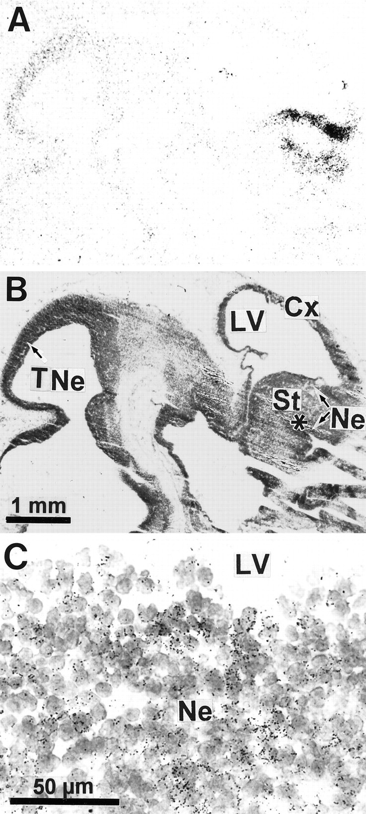
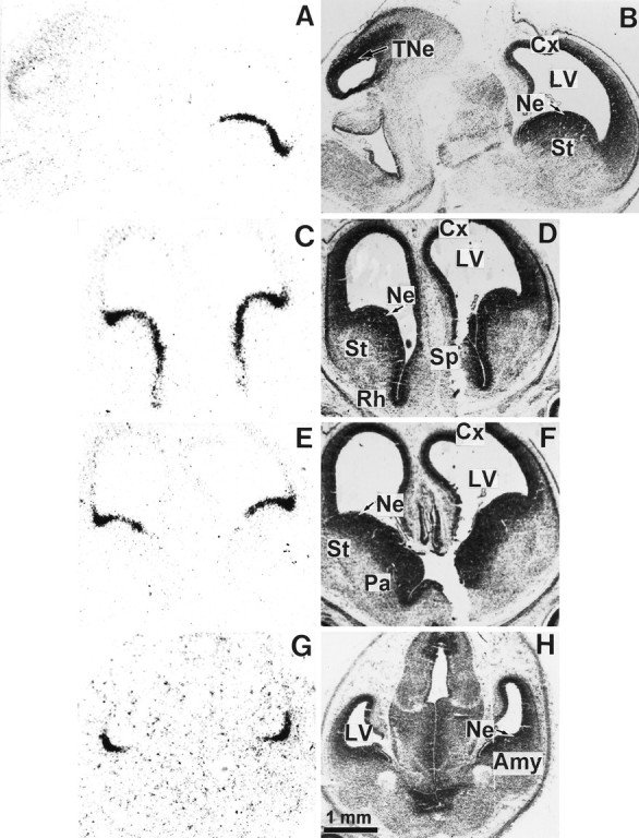
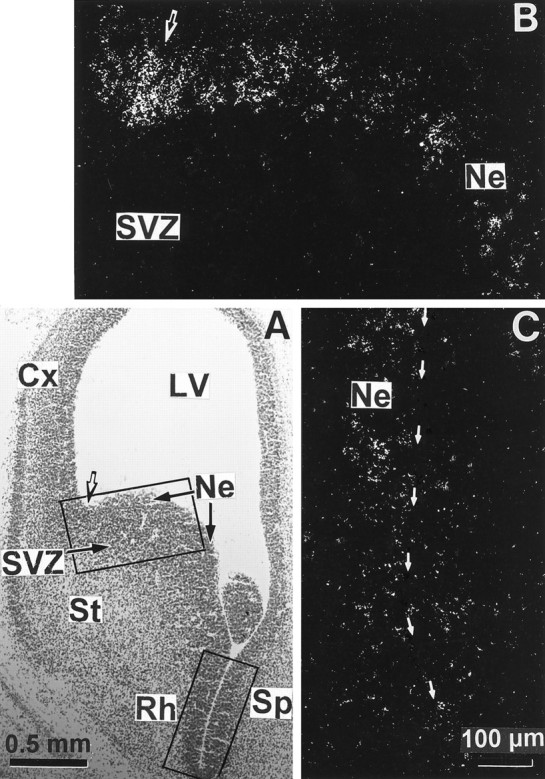
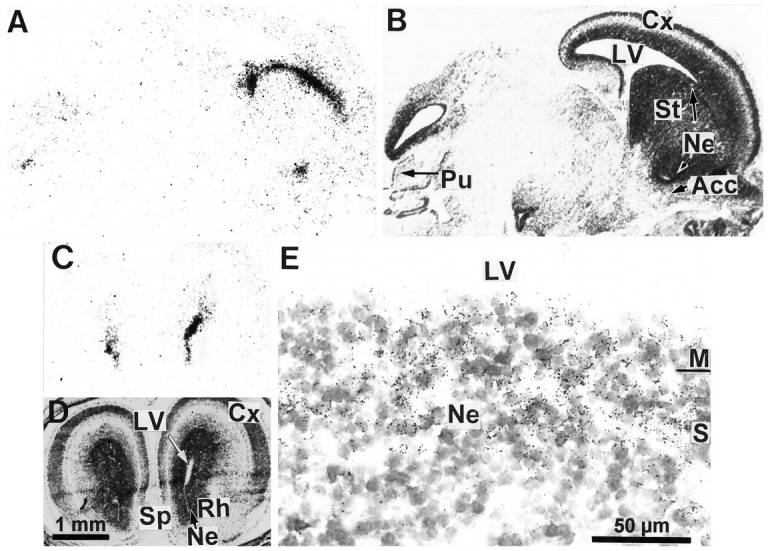
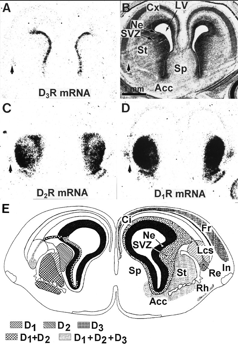
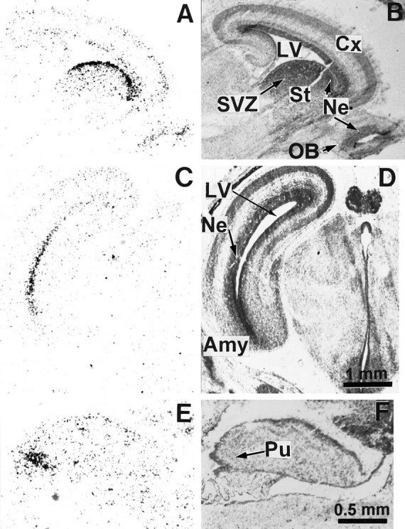
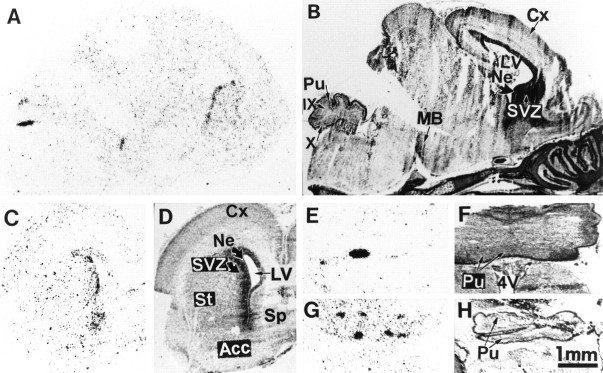
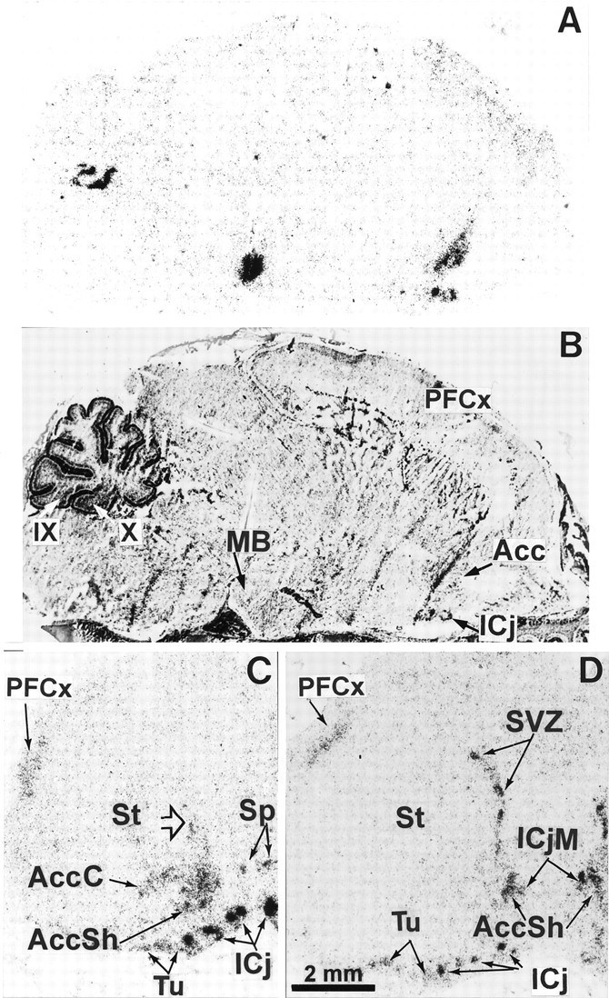
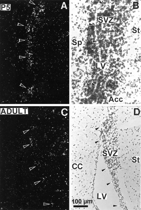
We studied by in situ hybridization histochemistry the expression of D3 receptor (D3R) mRNA at various stages of rat brain development. The first expression of D3R mRNA was detected at embryonic day 14 (E14) in the striatal and rhinencephalic neuroepithelia and throughout the tectal neuroepithelium. From E16 to E19 D3R mRNA expression extended along a rostrocaudal axis to additional proliferative ventricular zones of the basal forebrain, including the neuroepithelia of the olfactory bulb, nucleus accumbens, septum, and amygdala, whereas D1 and D2 receptor (D1R and D2R) mRNAs were expressed predominantly by migrating neuroblasts and/or differentiating striatal neurons. Only a few neuroblasts, migrating in the lateral cortical stream or developing as cerebellar Purkinje cells, expressed D3R mRNA from E18. At birth D3R expression mRNA appeared in differentiating neuronal fields of the nucleus accumbens and medial mamillary body primordia and on P5 reached a distribution similar to that found in adult. In addition, a transient upregulation was detected on P5 in the medial mamillary bodies, parietofrontal cortex, and olfactory tubercle. In the adult brain D3R gene expression continued in the striatal proliferative subventricular zone. The late expression D3R mRNA in neurons, after achievement of dopamine innervation, supports the existence of a regulating factor released from dopamine neurons, as suggested by denervation studies in the adult. The sustained and abundant D3R gene expression, predominantly in germinative neuroepithelial zones actively involved in neurogenesis of most basal forebrain structures, supports the hypothesis of a neurogenetic but minor morphogenetic modulatory role for the D3R during CNS development.


Figures

Fig. 1.
Fig. 1.
Expression of D3R mRNA in the brain of the 14 d embryo. A, Film autoradiographic hybridization signals from a parasagittal section hybridized with a D3R mRNA antisense probe. Hybridization signals are present only in neuroepithelia of striatum (St), rhinencephalon (indicated by asterisk), and tectum (TNe). Ct, Cortex. B, Structure locations are shown on the same section counterstained with Mayer’s hemalum solution and photographed with bright-field illumination. C, Photomicrograph of an emulsion-dipped section with bright-field illumination at the level of striatal neuroepithelium lining the lateral ventricle (LV), showing groups of labeled cells identified by clusters of dark silver grains overlying individual nuclei in the neuroepithelium (Ne).
Fig. 2.
Fig. 2.
Expression of D3R mRNA in the brain of the 16 d embryo. The left panel shows film autoradiograms from a parasagittal section (A) and three coronal serial sections (C, E,G) hybridized with the D3R mRNA probe. TheC, E, and G sections are ordered from rostral to caudal forebrain levels. The right panel shows the same sections counterstained with Mayer’s hemalum and photographed in bright-field illumination. The expression of D3R gene transcripts in the basal forebrain neuroepithelium, continuous from rostral striatum (St) to amygdala (Amy), is interrupted remarkably at the pallidum level (Pa). Cx, Cortex;LV, lateral ventricle; Ne, neuroepithelium; Rh, rhinencephalon; Sp, septum; TNe, tectal neuroepithelium.
Fig. 3.
Fig. 3.
High magnifications of D3R mRNA hybridization signals in basal forebrain neuroepithelium of the 16 d embryo. The top and bottom rectangles in a bright-field photomicrograph from a coronal section counterstained with Mayer’s hemalum (A) delimit the fields shown inB and C, respectively. B,C, Dark-field photomicrographs taken from an emulsion-dipped section through the rostral striatal anlage that show the labeling in the striatal (St), rhinencephalic (Rh), and septal (Sp) neuroepithelia bordering the ventral horn of the lateral ventricle (LV), indicated by thin arrows inC, whereas the subventricular zone (SVZ) and the differentiating field of striatum are labeled only scarcely. The strongest signal is present approximately at the limit between striatal and cortical neuroepithelia in A, as determined by the dorsolateral corner of the lateral ventricle (indicated bythick arrows in A and B).Cx, Cortex; Ne, neuroepithelium.
Fig. 4.
Fig. 4.
Expression of D3R mRNA in the basal forebrain and cerebellar primordia of the 18 d embryo. Autoradiograms of parasagittal (A) and coronal (C) sections hybridized with the D3R mRNA probe show signals in neuroepithelia (Ne) of striatum (St), nucleus accumbens (Acc), rhinencephalon (Rh), septum (Sp), and in the more caudal Purkinje cell layer (Pu) of cerebellar primordium. B, D, Corresponding sections counterstained with Mayer’s hemalum. High magnification of the striatal ventricular zone under bright-field illumination (E) illustrates that labeling is restricted to clusters of labeled cells present through the synthetic (S) and mitotic (M) neuroepithelial zones, delimiting a continuous periventricular band 100 μm thick. Cx, Cortex; LV, lateral ventricle; SVZ, subventricular zone.
Fig. 5.
Fig. 5.
Compared expressions of D1R, D2R, and D3R mRNAs in the forebrain of the 18 d embryo. Shown are film autoradiograms of adjacent coronal sections at a rostral level of the developing striatum, hybridized with probes specific for either D3R (A), D2R (C), or D1R (D) mRNAs. B, Bright-field photomicrograph of section A, counterstained with the Mayer’s hemalum. Cells in the reservoir of lateral cortical stream (indicated by arrows) express the three receptor subtype mRNAs (A, C, D). The cortical neuroepithelium and cortical plate (Cx) are labeled by the D1R and D3R, but not D2R, mRNA probes. D1R, D2R, and D3R mRNAs are expressed in different striatal compartments, as shown inE, which represents a schematic coronal section at the same level and developmental stage, taken from the atlas by Bayer and Altman (1995) in which the right cerebral hemisphere displays the structures (Acc, nucleus accumbens; Ci, cingulate cortex; Fr, frontal cortex; In, insulate cortex; Lcs, lateral cortical stream;Re, reservoir; Rh, rhinencephalon;Sp, septum; St, striatum) and the left hemisphere displays the respective distributions of D1R, D2R, and D3R mRNAs. In the striatum D1R mRNA is present in the differentiation field (St), D2R mRNA in the subventricular zone (SVZ) and in the differentiation field, and D3R mRNA in the neuroepithelium (Ne).
Fig. 6.
Fig. 6.
Expression of D3R mRNA in the brain of the 19 d embryo. The left panel shows film autoradiograms from parasagittal (A, E) and coronal (C) sections hybridized with the D3R mRNA probe. The right panels show corresponding sections counterstained with Mayer’s hemalum and photographed in bright-field illumination. Hybridization signals are detected at high level in several periventricular neuroepithelial regions (Ne) of the basal forebrain, including the striatum (St in A), amygdala (Amy in C), and in a group of Purkinje cells (Pu in E). Low expression of D3R is seen in the ventricular zone of the cortex and cortical plate (Cx), olfactory bulb (OB), and subventricular zone (SVZ) of the striatum inA. LV, Lateral ventricle.
Fig. 7.
Fig. 7.
In situ hybridization of D3R mRNA in the brain of a newborn pup. A,C, E, and G are autoradiograms of parasagittal (A) and coronal (C, E, G) sections hybridized with the D3R probe; B,D, F, and H are corresponding sections counterstained with Mayer’s hemalum. D3R mRNA is seen in the residual neuroepithelium (Ne) and the adjacent subventricular zone (SVZ) of the striatum (St), nucleus accumbens (Acc), and septum (Sp) as well as in the medial mamillary body (MB) and as patches in the Purkinje cell layer (Pu) of lobules 9 (IX) and 10 (X) in the cerebellar cortex. Cx, Cerebral cortex;LV, lateral ventricle; 4V, fourth ventricle.
Fig. 8.
Fig. 8.
Expression of D3R mRNA in the brain of the 5 d pup. Shown are autoradiograms of parasagittal (A) and coronal [rostral (C) and caudal (D) level of nucleus accumbens] sections hybridized with the D3R probe. B corresponds to sectionA, counterstained with Mayer’s hemalum. High expression of transcripts is observed in the major (ICjM) and minor (ICj) islands of Calleja, nucleus accumbens shell (AccSh), medial mamillary body (MB), patches in the subventricular zone (SVZ) of striatum (St), and Purkinje cell layer of the cerebellar lobules 9 (IX) and 10 (X), whereas moderate signals are found in the core of nucleus accumbens (AccC), ventromedial region of the striatum (arrowhead), septum (Sp), parietofrontal cortex (PFCx), and olfactory tubercle (Tu).
Fig. 9.
Fig. 9.
Expression of D3R gene transcripts in the subventricular zone of striatum in brains from a 5 d pup (A, B) or adult (C,D). Photomicrographs under dark-field illumination (A, C) show labeling in the striatal subventricular zone (SVZ) lining the lateral ventricle (LV), as indicated by arrowheads. B, D, Corresponding sections counterstained with Mayer’s hemalum. Acc, Nucleus accumbens;CC, corpus callosum; Sp, septum;St, striatum.

References

    1. Altman J, Bayer SA. Embryonic development of the rat cerebellum. III. Regional differences in the time of origin, migration, and settling of Purkinje cells. J Comp Neurol. 1985;231:42–65. - PubMed
    1. Altman J, Bayer SA. Vertical compartmentation and cellular transformations in the germinal matrices of the embryonic rat cerebral cortex. Exp Neurol. 1990;107:23–35. - PubMed
    1. Arenander AT, De Vellis J. Development of the nervous system. In: Siegel GJ, editor. Basic neurochemistry: molecular, cellular, and medical aspects. Raven; New York: 1994. pp. 573–606.
    1. Bayer SA, Altman J. Cell migration in the developing neocortex. In: Bayer SA, Altman J, editors. Neocortical development. Raven; New York: 1991. pp. 116–127.
    1. Bayer SA, Altman J. Principles of neurogenesis, neuronal migration, and neuronal circuit formation. In: Paxinos G, editor. The rat nervous system. Academic; New York: 1995. pp. 1079–1098.

Publication types

LinkOut - more resources