

The fusiform face area: a module in human extrastriate cortex specialized for face perception
- PMID: 9151747
- PMCID: PMC6573547
- DOI: 10.1523/JNEUROSCI.17-11-04302.1997
The fusiform face area: a module in human extrastriate cortex specialized for face perception
Abstract
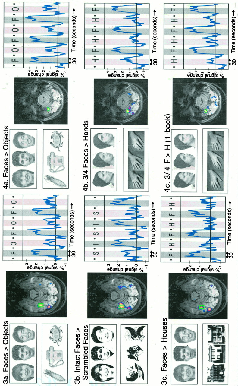
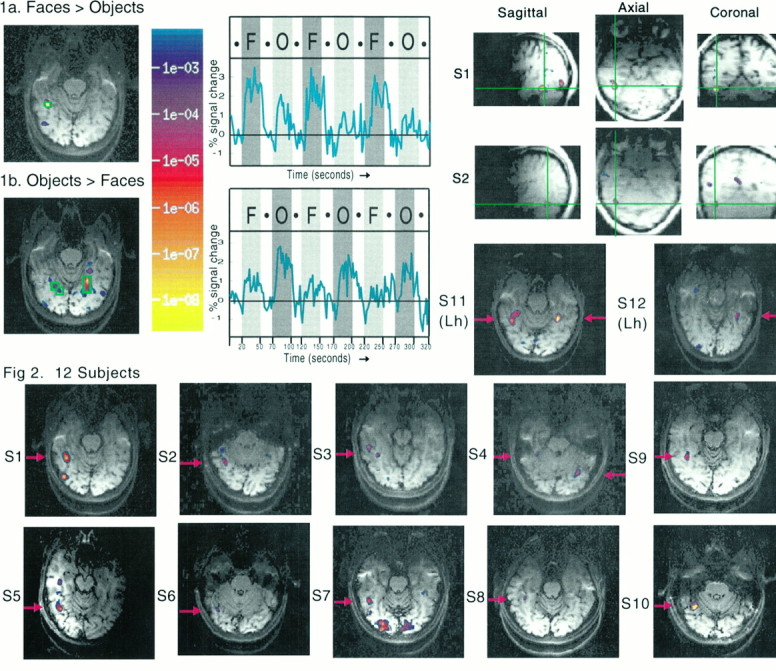
Using functional magnetic resonance imaging (fMRI), we found an area in the fusiform gyrus in 12 of the 15 subjects tested that was significantly more active when the subjects viewed faces than when they viewed assorted common objects. This face activation was used to define a specific region of interest individually for each subject, within which several new tests of face specificity were run. In each of five subjects tested, the predefined candidate "face area" also responded significantly more strongly to passive viewing of (1) intact than scrambled two-tone faces, (2) full front-view face photos than front-view photos of houses, and (in a different set of five subjects) (3) three-quarter-view face photos (with hair concealed) than photos of human hands; it also responded more strongly during (4) a consecutive matching task performed on three-quarter-view faces versus hands. Our technique of running multiple tests applied to the same region defined functionally within individual subjects provides a solution to two common problems in functional imaging: (1) the requirement to correct for multiple statistical comparisons and (2) the inevitable ambiguity in the interpretation of any study in which only two or three conditions are compared. Our data allow us to reject alternative accounts of the function of the fusiform face area (area "FF") that appeal to visual attention, subordinate-level classification, or general processing of any animate or human forms, demonstrating that this region is selectively involved in the perception of faces.
Figures
References
-
- Allison T, Ginter H, McCarthy G, Nobre AC, Puce A, Belger A. Face recognition in human extrastriate cortex. J Neurophysiol. 1994;71:821–825. - PubMed
-
- Behrmann M, Winocur G, Moscovitch M. Dissociation between mental imagery and object recognition in a brain-damaged patient. Nature. 1992;359:636–637. - PubMed
-
- Breiter HC, Etcoff NL, Whalen PJ, Kennedy WA, Rauch SL, Buckner RL, Strauss MM, Hyman SE, Rosen BR. Response and habituation of the human amygdala during visual processing of facial expression. Neuron. 1996;17:875–887. - PubMed
-
- Bruce V, Doyle T, Dench N, Burton M. Remembering facial configurations. Cognition. 1991;38:109–144. - PubMed
Publication types
MeSH terms
LinkOut - more resources
Full Text Sources
Other Literature Sources