. 1997 Jun 1;17(11):4367-81.
doi: 10.1523/JNEUROSCI.17-11-04367.1997.

Transient changes in flocculonodular lobe protein kinase C expression during vestibular compensation

Affiliations

Transient changes in flocculonodular lobe protein kinase C expression during vestibular compensation

M M Goto et al. J Neurosci. .

Abstract

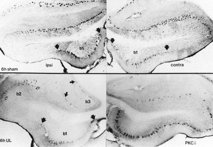
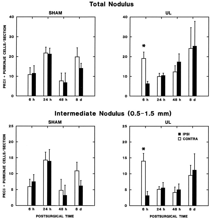
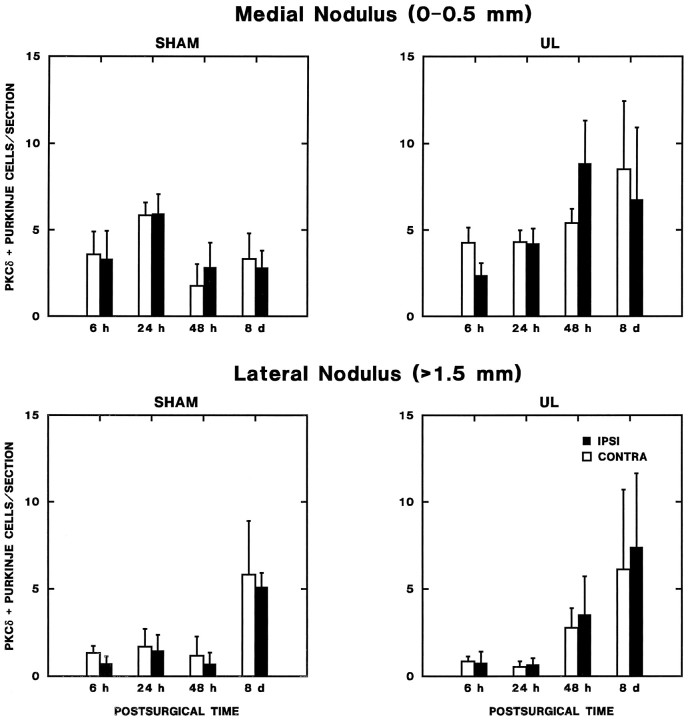
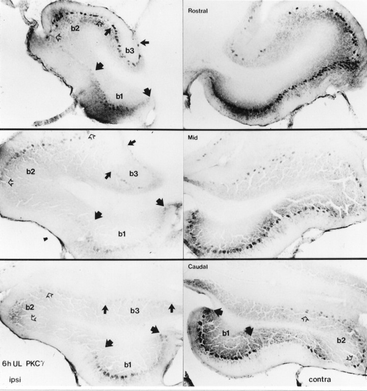
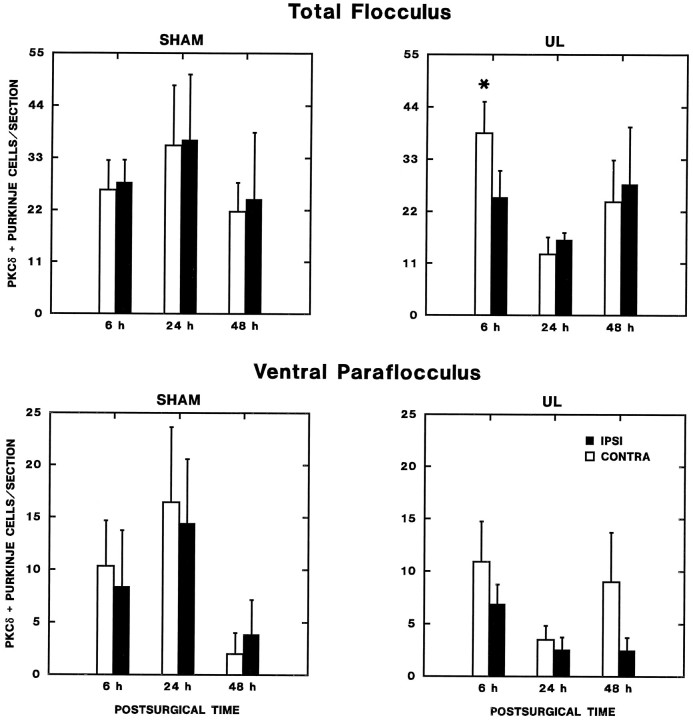
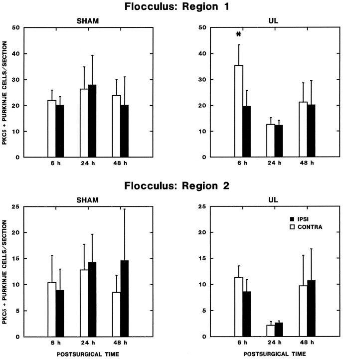
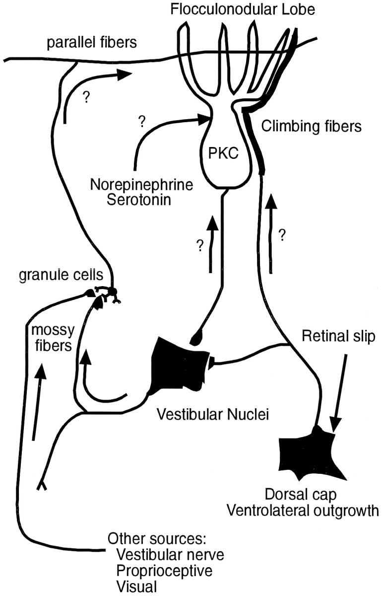
Protein kinase C (PKC) is a family of intracellular signal transduction enzymes, comprising isoforms that vary in sensitivity to calcium, arachidonic acid, and diacylglycerol. PKC isoforms alpha, gamma, and delta are expressed by cerebellar Purkinje cells and neurons in the cerebellar nuclei and vestibular nuclei of the Long-Evans rat. In control rats, these PKCs are distributed symmetrically in the flocculonodular-lobe Purkinje cells. Behavioral recovery from vestibular dysfunction produced by unilateral labyrinthectomy (UL) is accompanied by asymmetric expression of PKC isoforms in these regions within 6 hr after UL. These expression changes were localized within parasagittal regions of the flocculus and nodulus. The distribution of PKCalpha, -gamma, and -delta were identical, suggesting that they are coregulated in cerebellar Purkinje cells during this early compensatory period. The pattern of Purkinje cell PKC expression returned to the control, symmetric distribution within 24 hr after UL. It is hypothesized that these regional changes in Purkinje cell PKC expression are an early intracellular signal contributing to vestibular compensation. In particular, regulation of PKC expression may contribute to changes in the efficacy of cerebellar synaptic plasticity during the acute post-UL period.


Figures

Fig. 1.
Fig. 1.
Histological verification of unilateral surgical labyrinthectomies. Photomicrographs of horizontal sections through the unoperated (A) and operated (B) ears of case 9311 are shown, with rostral toward the left. The medial aspect of the section is oriented upward inA and downward in B andC. Note the intact sacculus in A(black arrow) and the complete ablation of the sacculus on the operated side (B, black arrow).Open arrows show the unaffected Scarpa’s ganglia on both the unoperated (A) and operated (B) sides. The Scarpa’s ganglion from the operated side is shown at higher magnification in C. Scale bars: A,B, 500 μm; C, 100 μm.
Fig. 2.
Fig. 2.
Western immunoblot of PKCδ from the cerebellar posterior lobe vermis of 6 hr post-sham-operated (S) and unilateral labyrinthectomy (UL) Long–Evans rats. PKCδ migrates as a 74 and 76 kDa doublet representing its two phosphorylation states. There are no changes in migration distances between sham and UL samples membrane (M) or cytoplasmic (C) fractions. This demonstrates antibody specificity and suggests that the electrophoretic motility of PKCδ does not change within 6 hr of labyrinthectomy. Reprobing of the blot showed no change in migration distances for PKCα or -γ. Molecular weight markers are shown to the left.
Fig. 3.
Fig. 3.
Cellular distribution of PKC immunoreactivity in the flocculus (A, C) and nodulus (B, D). A, PKCδ immunoreactivity in the flocculus was observed in somata and dendrites of many Purkinje cells and in somata of some molecular layer interneurons (open arrows). The immunopositive interneurons were observed in bands along the ventromedial and ventrolateral margins of the flocculus. Erythrocytes are indicated bysmall black arrows. B, PKCδ-immunopositive cells are shown in a tangential section through lobule Xa of the nodulus. Note the intense immunoreactivity of Purkinje cell somata and proximal dendrites. Weakly immunopositive molecular layer interneurons were observed rarely (open arrow).Small black arrows indicate erythrocytes.C, Higher magnification photomicrograph of somatodendritic staining of flocculus Purkinje cells from a band showing no immunopositive molecular layer interneurons.D, High magnification view of PKCα immunopositive Purkinje cells (arrows) from a tangential section through the Purkinje cell layer of lobule Xa. Note the intense immunoreactivity associated with the nuclear region and the weaker reaction in the somata and proximal dendrites.
Fig. 4.
Fig. 4.
Distribution of PKCδ expression in transverse sections through cerebellar lobules IX (uvula) and X (nodulus) 6 hr postoperatively. These photomicrographs at two comparable levels illustrate results from lobules IXc and Xa from a sham-operated (case 9330) and a UL (case 9329) rat. Expression in the cerebellar cortex was limited to Purkinje cell somata and dendrites, but not all Purkinje cells showed PKCδ immunoreactivity. Six hours after UL, PKCδ-positive Purkinje cells were distributed in a strikingly asymmetric pattern, with markedly increased expression in an intermediate band in the nodulus contralateral to the labyrinthectomy (solid arrows). By contrast, 6 hr after a sham operation, Purkinje cells expressing PKCδ were distributed in a more symmetric pattern across the nodulus and uvula, with relatively few immunopositive cells in the intermediate region. PKCδ-immunoreactivity in the granular layer (asterisks) and interneurons in the molecular layer (open arrows) showed no consistent patterns across animals. The pattern of PKCα and -γ immunopositive Purkinje cell somata was identical to the distribution of PKCδ immunoreactivity.
Fig. 8.
Fig. 8.
Distribution of PKCδ expression in sections through Long–Evans rat cerebellar flocculus. As in the nodulus and uvula, immunoreactivity is restricted to the Purkinje cells of the cerebellar cortex. The flocculus ipsilateral to the operation is shown on the left side and the contralateral flocculus is shown on the right side of the figure. Six hour, post-sham-operated rat flocculi (top panels) show a variable but consistently symmetric pattern of Purkinje cell immunoreactivity along the dorsal and ventromedial (b1, demarcated by wide black arrows) surfaces. Six hour, post-UL flocculi (lower panels) show the ventromedial distribution of expression as in the sham, with the appearance of bands of expression along the lateral crest of the ipsilateral flocculus (b2, open arrows) and the border with paraflocculus (b3, narrow black arrows). The contralateral flocculus shows a broader area of distribution than ipsilaterally, with expression between areas b1 andb2, and b2 and the lateral margin of b3. Contralaterally, areab3 shows only a sparse distribution of immunoreactive Purkinje cells. Immunoreactivity for PKCα and -γ shows an identical distribution.
Fig. 5.
Fig. 5.
Density histograms of PKCδ immunoreactive Purkinje cells across the rat nodulus and uvula. These data were obtained from every sixth section through these regions. Folia of the nodulus and uvula are indicated along the x-axis. The midline of the cerebellar cortex is indicated with a “0” along they-axis. Positive distances represent millimeters from the midline contralateral to the side of operation and negative values the ipsilateral side. The vertical axis represents immunoreactive Purkinje cell counts in 250 μm increments. Each histogram represents a different rat; the case number is given on each histogram. Density histograms of 6 hr post-sham-operation show a symmetric distribution of immunoreactive Purkinje cells near the midline that is consistent between rat cerebella and levels of PKC expression. In 6 hr post-UL rat cerebella, the distribution was asymmetric in all rats.
Fig. 6.
Fig. 6.
Zonal changes in lobule Xa (nodulus) PKCδ expression during vestibular compensation. The number of PKCδ-immunopositive Purkinje cells per section from every sixth section through the lobule Xa of the nodulus ipsilateral and contralateral to operations is plotted as a function of postoperative time. Separate graphs are shown for the entire nodulus (total nodulus, top panels) and for the intermediate zone (0.5–1 mm lateral to the midline, lower panels). Results of repeated-measures ANOVA and post hoc comparisons are presented in the text. The asterisks indicate a significant elevation in PKC expression on the side contralateral to the operation in the 6 hr UL rats (Newman–Keuls test;p < 0.05 vs ipsilateral side and sham-operated controls at 6 hr).
Fig. 7.
Fig. 7.
Zonal changes in lobule Xa (nodulus) PKCδ expression during vestibular compensation. The number of PKCδ-immunopositive Purkinje cells per section from every sixth section through the lobule Xa of the nodulus ipsilateral and contralateral to operations is plotted as a function of postoperative time. Separate graphs are shown for the medial nodulus (0–0.5 mm from the midline) and the lateral nodulus (>1.5 mm from the midline). No significant effects were observed in the medial nodulus. In the lateral nodulus, there was a bilaterally symmetric increase in the number of PKC-immunopositive Purkinje cells in both sham and UL groups on the eighth postoperative day (see text).
Fig. 9.
Fig. 9.
Rostrocaudal distribution of PKCγ expression in the Long–Evans rat flocculus. The symmetric banding along the dorsal and ventromedial (b1, demarcated by wide black arrows) floccular surfaces in the 6 hr sham-operated rat runs caudorostrally. In the 6 hr post-UL flocculi, the broader contralateral banding between b1 and b2 (demarcated byopen arrows), and b2 and the lateral margin of b3 (demarcated by narrow black arrows) runs to merge on the dorsal surface of the rostral pole. The sparser band of immunoreactivity in contralateralb3 also runs caudorostrally. PKCα and -δ show the same distribution of expression.
Fig. 10.
Fig. 10.
Changes in flocculus and ventral paraflocculus Purkinje cell PKCδ expression. The number of PKCδ-immunopositive Purkinje cells per section (from every sixth section through the flocculus and ventral paraflocculus) ipsilateral and contralateral to operations is plotted as a function of postoperative time. Separate graphs are shown for the total flocculus (top panels) and ventral paraflocculus. In the flocculus, there was a statistically significant increase in PKC expression contralateral to the lesion in the 6 hr UL group (Newman–Keuls test; p < 0.01 compared with the ipsilateral side and both sides of the 6 hr sham-operated group). No significant effects were observed in the ventral paraflocculus.
Fig. 11.
Fig. 11.
Transient regional changes in flocculus Purkinje cell expression of PKCδ. The number of PKCδ-immunopositive Purkinje cells per section from every sixth section through the intermediate aspect of flocculus is plotted as a function of postoperative time. Increased expression was observed contralaterally 6 hr after UL (Newman–Keuls test; p < 0.01) in region 1, which included the entire ventral surface and intermediate third of the dorsal surface of the nodulus. No significant effects were observed in region 2, the lateral and medial thirds of the dorsal surface of the flocculus.
Fig. 12.
Fig. 12.
Schematic diagram of neural circuits that may influence PKC expression during vestibular compensation. Two potential substrates for the parasagittal distribution of increased PKC expression in the flocculonodular lobe are (1) an anterograde influence of climbing fiber projections from the dorsal cap and ventrolateral outgrowth and (2) a retrograde influence via zonal projections of Purkinje cells to the vestibular nuclei. A contribution of mossy fiber–granule cell-parallel fiber and monoaminergic projections, however, cannot be excluded.

References

    1. Azzi A, Boscoboinik D, Hensey C. The protein kinase C family. Eur J Biochem. 1992;208:547–557. - PubMed
    1. Balaban CD. Olivovestibular and cerebellovestibular connections in albino rabbits. Neuroscience. 1984;12:129–149. - PubMed
    1. Balaban CD. Distribution of inferior olivary projections to the vestibular nuclei of albino rabbits. Neuroscience. 1988;24:119–134. - PubMed
    1. Balaban CD, Henry RT. Zonal organization of olivo-nodulus projections in albino rabbits. Neurosci Res. 1988;5:409–423. - PubMed
    1. Balaban CD, Billingsley ML, Kincaid RL. Evidence for transsynaptic regulation of calmodulin-dependent cyclic nucleotide phosphodiesterase in cerebellar Purkinje cells. J Neurosci. 1989;9:2374–2381. - PMC - PubMed

Publication types

Substances

LinkOut - more resources