. 1997 May 5;185(9):1595-604.
doi: 10.1084/jem.185.9.1595.

Human macrophage-derived chemokine (MDC), a novel chemoattractant for monocytes, monocyte-derived dendritic cells, and natural killer cells

Affiliations

Human macrophage-derived chemokine (MDC), a novel chemoattractant for monocytes, monocyte-derived dendritic cells, and natural killer cells

R Godiska et al. J Exp Med. .

Abstract

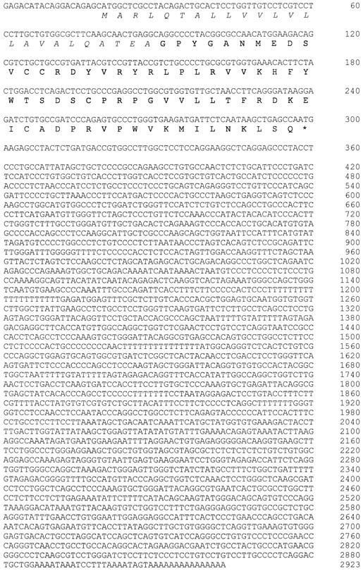
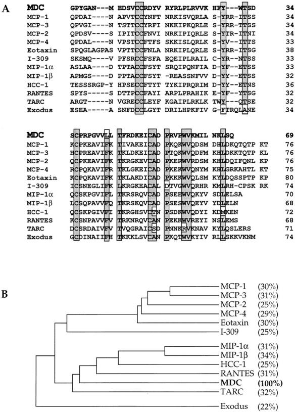
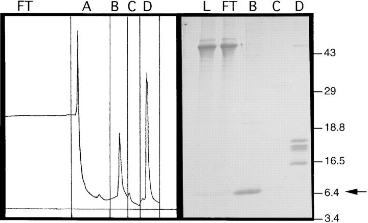
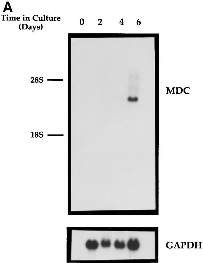
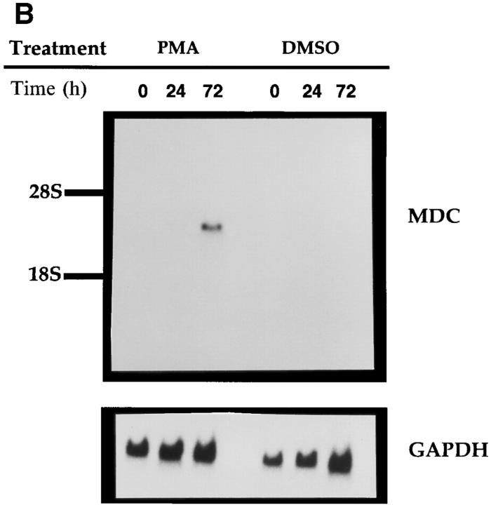
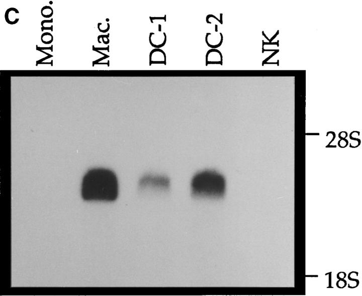
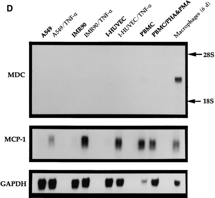
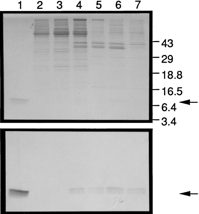
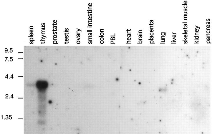
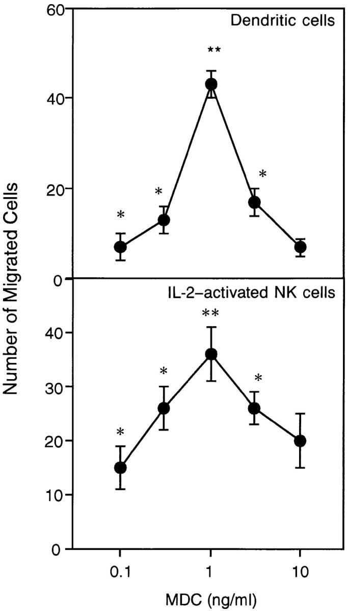
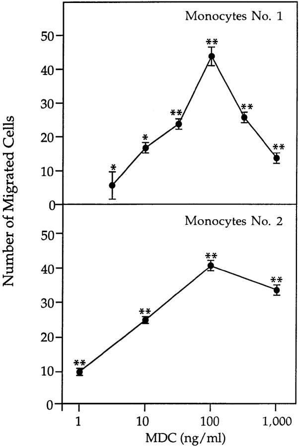
A cDNA encoding a novel human chemokine was isolated by random sequencing of cDNA clones from human monocyte-derived macrophages. This protein has been termed macrophage-derived chemokine (MDC) because it appears to be synthesized specifically by cells of the macrophage lineage. MDC has the four-cysteine motif and other highly conserved residues characteristic of CC chemokines, but it shares <35% identity with any of the known chemokines. Recombinant MDC was expressed in Chinese hamster ovary cells and purified by heparin-Sepharose chromatography. NH2-terminal sequencing and mass spectrophotometry were used to verify the NH2 terminus and molecular mass of recombinant MDC (8,081 dalton). In microchamber migration assays, monocyte-derived dendritic cells and IL-2-activated natural killer cells migrated to MDC in a dose-dependent manner, with a maximal chemotactic response at 1 ng/ml. Freshly isolated monocytes also migrated toward MDC, but with a peak response at 100 ng/ml MDC. Northern analyses indicated MDC is highly expressed in macrophages and in monocyte-derived dendritic cells, but not in monocytes, natural killer cells, or several cell lines of epithelial, endothelial, or fibroblast origin. High expression was also detected in normal thymus and less expression in lung and spleen. Unlike most other CC chemokines, MDC is encoded on human chromosome 16. MDC is thus a unique member of the CC chemokine family that may play a fundamental role in the function of dendritic cells, natural killer cells, and monocytes.


Figures

Figure 1
Figure 1
Sequence and deduced translation of the MDC cDNA. The leader sequence is in italics; the mature protein is in bold type. The mature NH2 terminus was confirmed by amino acid sequencing of recombinant MDC produced in CHO cells (GPYGANMEDSV . . .). Alu repeats (68) are present in the following positions: bases 1204–1482, 1530–1805, and 2335–2443. The cDNA sequence of MDC has been deposited in EMBL/GenBank/DDBJ under the accession number U83171.
Figure 2
Figure 2
Comparison of the mature form of human MDC with the known human CC chemokines. (A) Direct comparison of amino acid sequences. Residues conserved in at least 10 of the 12 sequences are shaded; the exceptions are boxed and unshaded. Dashes are inserted to optimize alignment of the sequences. (B) Dendrogram analysis illustrating the similarity of the mature forms of the CC chemokines. Percent identity with MDC is indicated in parentheses.
Figure 3
Figure 3
Purification of recombinant MDC from CHO cells. A PCR fragment containing bases 1–403 of the MDC cDNA was subcloned into a mammalian expression vector driven by the CMV promoter and transfected into CHO cells. Culture supernatant from an individual transfected clone was harvested 4 d after cells reached confluency. MDC in the media was concentrated and purified by binding to a heparin-Sepharose column in 0.35 M NaCl. The left panel shows the column chromatogram. Fractions collected were flowthrough (FT), 0.35 M NaCl wash (A), 0.7 M NaCl elution (B), repeat of 0.7 M NaCl elution (C), and 1.5 M NaCl elution (D). The right panel shows SDS-PAGE analysis (18% Tris glycine) of the supernatant loaded onto the column (L) and fractions FT, B, C, and D. MDC eluted in fraction B (arrow). The gel was transferred to polyvinylidene difluoride membrane, and the MDC band was excised for NH2-terminal sequencing.
Figure 4
Figure 4
Northern analysis of MDC mRNA expression in human cells. Migration of MDC, 28S RNA, and 18S RNA is indicated. (A) Peripheral blood monocytes were allowed to differentiate into macrophages by incubation on plastic tissue culture dishes for 6 d (41). Total RNA was isolated from individual dishes on the indicated days, fractionated through a formaldehyde agarose gel, blotted, and probed with the radiolabeled MDC cDNA. The blot was subsequently probed for GAPDH message. (B) Expression of MDC in differentiated HL-60 cells. Cells were treated with PMA to induce differentiation to monocytic cells or with DMSO to induce granulocytic cells. MDC expression was analyzed by Northern blotting as in A. (C) Northern blot of MDC expression in human peripheral blood monocytes (Mono.), macrophages derived from these monocytes (Mac.), dendritic cells derived from PBMC of two different donors (DC-1 and DC-2), and natural killer cells derived from PBMC (NK). (D) Expression of MDC in cell cultures. Lung epithelial cell line A549, lung fibroblast line IMR90, and I-HUVEC without and with TNF-α stimulation; PBMC without and with stimulation by PHA and PMA; and monocyte derived macrophages after 6 d in culture. After probing with MDC, the filter was stripped and probed sequentially with MCP-1 and GAPDH.
Figure 4
Figure 4
Northern analysis of MDC mRNA expression in human cells. Migration of MDC, 28S RNA, and 18S RNA is indicated. (A) Peripheral blood monocytes were allowed to differentiate into macrophages by incubation on plastic tissue culture dishes for 6 d (41). Total RNA was isolated from individual dishes on the indicated days, fractionated through a formaldehyde agarose gel, blotted, and probed with the radiolabeled MDC cDNA. The blot was subsequently probed for GAPDH message. (B) Expression of MDC in differentiated HL-60 cells. Cells were treated with PMA to induce differentiation to monocytic cells or with DMSO to induce granulocytic cells. MDC expression was analyzed by Northern blotting as in A. (C) Northern blot of MDC expression in human peripheral blood monocytes (Mono.), macrophages derived from these monocytes (Mac.), dendritic cells derived from PBMC of two different donors (DC-1 and DC-2), and natural killer cells derived from PBMC (NK). (D) Expression of MDC in cell cultures. Lung epithelial cell line A549, lung fibroblast line IMR90, and I-HUVEC without and with TNF-α stimulation; PBMC without and with stimulation by PHA and PMA; and monocyte derived macrophages after 6 d in culture. After probing with MDC, the filter was stripped and probed sequentially with MCP-1 and GAPDH.
Figure 5
Figure 5
MDC protein expression in human monocyte–derived macrophages and unstimulated epithelial cell lines. Monocytes were differentiated into macrophages by incubation on tissue culture plastic for 6 d. LDL or oxidized LDL was then added for an additional 3 d. Culture supernatants from these macrophages or from epithelial lines were passed over heparin– Sepharose columns, and MDC was eluted with 0.6 M NaCl, fractionated by SDS-PAGE, and reacted with an mAb raised against recombinant MDC produced in bacteria. (Top) The Coomassie stained gel of the heparin– Sepharose elutates. (Bottom) The Western blot. Lanes 1, CHO-derived MDC; 2, A549 epithelial cell; 3, T84 epithelial cell; 4, macrophage, day 6; 5, macrophage, day 9; 6, macrophage plus LDL, day 9; 7, macrophage plus oxidized LDL, day 9. Arrows indicate the migration of MDC.
Figure 6
Figure 6
Expression of MDC mRNA in normal human tissues. A multiple tissue Northern blot was probed with the MDC cDNA sequence and washed at high stringency (0.2× SSC, 50°C). The migration of RNA size markers is indicated in kb.
Figure 7
Figure 7
Chemotaxis of monocyte-derived dendritic cells and IL-2–activated natural killer cells induced by MDC. (Top) PBMC were isolated by density gradient centrifugation and depleted of CD19+ and CD2+ cells by antibody-coated magnetic beads. Dendritic cells were obtained by culturing the remaining cells for 6 to 8 d in RPMI containing containing 10% FCS, 50 ng/ml GM-CSF, and 20 ng/ ml IL-13. (Bottom) Natural killer cells were purified by discontinuous Percoll gradient centrifugation of monocyte-depleted PBMC, followed by negative selection of T cells by panning with an anti-CD6 mAb. Cells were cultured with an irradiated lymphoblastoid cell line in the presence of 250 U/ml IL-2. For chemotaxis assays, cells were added to the upper wells of a microchamber and medium (RPMI plus 1% FCS) with or without chemokine was added to the lower chamber. The figure shows representative experiments. Values represent the number of migrated cells (mean ± SE) after subtraction of basal migration (24 ± 3 for dendritic cells and 20 ± 6 for natural killer cells). *P <0.05), **P <0.01.
Figure 8
Figure 8
Chemotaxis of monocytes induced by MDC. Human PBMC were purified by density gradient centrifugation and resuspended in RPMI plus 1% FCS. Chemotaxis was assayed as described in Fig. 7; two independent experiments are illustrated. Under these conditions, only monocytes migrated through the filter. Basal migration of 15 ± 2 (top) and 16 ± 2 (bottom) was subtracted from the values shown. The response of these cells to 100 ng/ml MCP-3 was 42 ± 4 and to 10 nM fMLP was 54 ± 3.

References

    1. Oppenheim JJ. Overview of chemokines. Adv Exp Med Biol. 1993;351:183–186. - PubMed
    1. Baggiolini M, Dewald B, Moser B. Interleukin-8 and related chemotactic cytokines-CXC and CC chemokines. Adv Immunol. 1994;55:97–179. - PubMed
    1. Schall TJ. Biology of the RANTES/SIS cytokine family. Cytokine. 1991;3:165–183. - PubMed
    1. Eaves CJ, Cashman JD, Wolpe SD, Eaves AC. Unresponsiveness of primitive chronic myeloid leukemia cells to macrophage inflammatory protein 1 α, an inhibitor of primitive normal hematopoietic cells. Proc Natl Acad Sci USA. 1993;90:12015–12019. - PMC - PubMed
    1. Tashiro K, Tada H, Heilker R, Shirozu M, Nakano T, Honjo T. Signal sequence trap: a cloning strategy for secreted proteins and type I membrane proteins. Science (Wash DC) 1993;261:600–603. - PubMed

Publication types

MeSH terms

Associated data