. 1997 May 27;94(11):5525-30.
doi: 10.1073/pnas.94.11.5525.

Design of polydactyl zinc-finger proteins for unique addressing within complex genomes

Affiliations

Design of polydactyl zinc-finger proteins for unique addressing within complex genomes

Q Liu et al. Proc Natl Acad Sci U S A. .

Abstract

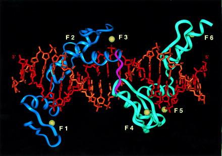
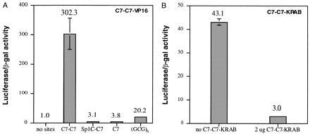
Zinc-finger proteins of the Cys2-His2 type represent a class of malleable DNA-binding proteins that may be selected to bind diverse sequences. Typically, zinc-finger proteins containing three zinc-finger domains, like the murine transcription factor Zif268 and the human transcription factor Sp1, bind nine contiguous base pairs. To create a class of proteins that would be generally applicable to target unique sites within complex genomes, we have utilized structure-based modeling to design a polypeptide linker that fuses two three-finger proteins. Two six-fingered proteins were created and demonstrated to bind 18 contiguous bp of DNA in a sequence-specific fashion. Expression of these proteins as fusions to activation or repression domains allows transcription to be specifically up- or down-modulated within human cells. Polydactyl zinc-finger proteins should be broadly applicable as genome-specific transcriptional switches in gene therapy strategies and the development of novel transgenic plants and animals.


Figures

Figure 1
Figure 1
Model of a six zinc-finger–DNA complex. The zinc fingers (numbered F1–F3 and F4–F6) are based on the structure of the three zinc-finger protein Zif268 (3). The six-finger protein was constructed by connecting F3 to F4 by building in the conserved linker sequence TGEKP, represented in magenta, with insightii. The guanine-rich strand, with which most of the base-specific contacts are made, is represented in red (3′-TGCGGGTGCGGCGGGTGCGA-5′). The zinc atoms are represented in yellow. The structure of the modeled linker is similar to that of the natural linker peptides between F1 and F2 (TGQKP) and between F2 and F3 (TGEKP), respectively (with an rmsd of 0.3 Å for backbone atoms). The modeled linker also retains the general position and hydrogen bond characteristics observed for the natural linkers.
Figure 2
Figure 2
Binding of the maltose-binding protein fusions (MBP)-C7-C7 and MBP-Sp1C-C7 with duplex DNA oligonucleotides containing various target sequences. (A) MBP-C7-C7 protein was used to shift the double-stranded DNA probes containing the target sequences listed on top of each panel [from left to right: C7-C7 site, Sp1C-C7 site, C7 site, and (GCG)6 site]. The protein concentration is given in nanomoles (nM) beneath each lane with a 2-fold serial dilution from left to right in each panel. (B) MBP-SP1C-C7 protein was titrated into gel shift reactions with probes containing target sequences (from left to right: Sp1C-C7 site, C7-C7 site, C7 site, and Sp1C site) as listed on top of each panel. The protein concentration is labeled in nM beneath each lane, with a 2-fold serial dilution from left to right in each panel.
Figure 3
Figure 3
DNaseI footprint of MBP-C7-C7 and MBP-Sp1C-C7. A 220-bp radiolabeled fragment containing the binding site for MBP-C7-C7 (lanes 1–3) or MBP-Sp1C-C7 (lanes 4–6) was incubated with either 20 μg/ml of BSA (lanes 2 and 4) or the cognate binding protein (300 nM, lanes 3 and 6) in 1× binding buffer for 30 min. DNaseI footprinting was then performed using the SureTrack Footprinting Kit (Pharmacia) according to the manufacturer’s instructions. Boxed region indicates the binding site sequence. Asterisk indicates the 3′-labeled strand. Lanes 1 and 4: G + A ladders.
Figure 4
Figure 4
Transcriptional regulation mediated by six-finger proteins in living cells. (A) HeLa cells were transiently transfected in triplicate with 2.5 μg of the indicated reporter plasmids and 2.5 μg C7-C7-VP16 expression plasmid. Luciferase activities were measured 48 hr later and normalized to the control β-galactosidase activity. The relative light units are given on top of each column (error bar = SD). (B) HeLa cells were transfected with 2.5 μg of the indicated reporter plasmids and either no C7-C7-KRAB expression or 1 μg of the C7-C7-KRAB expression plasmid by using LipofectAmine (GIBCO/BRL) as the transfection reagent. Luciferase activities were measured 48 hr later, and normalized to the control β-galactosidase activity. The relative light unit values were labeled on top of each column (error bar = SD).

Similar articles

Cited by

References

    1. Miller J, McLachlan A D, Klug A. EMBO J. 1985;4:1609–1614. - PMC - PubMed
    1. Berg J M, Shi Y. Science. 1996;271:1081–1085. - PubMed
    1. Pavletich N P, Pabo C O. Science. 1991;252:809–817. - PubMed
    1. Fairall L, Schwabe J W, Chapman L, Finch J T, Rhodes D. Nature (London) 1993;366:483–487. - PubMed
    1. Pavletich N P, Pabo C O. Science. 1993;261:1701–1707. - PubMed

Publication types

MeSH terms