Comparative Study
. 1997 May 27;94(11):5744-9.
doi: 10.1073/pnas.94.11.5744.

Characterization of the major histocompatibility complex class II binding site on LAG-3 protein

Affiliations
Comparative Study

Characterization of the major histocompatibility complex class II binding site on LAG-3 protein

B Huard et al. Proc Natl Acad Sci U S A. .

Abstract

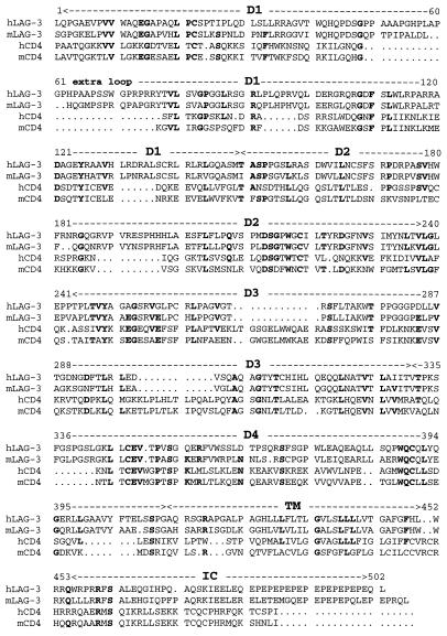
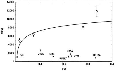
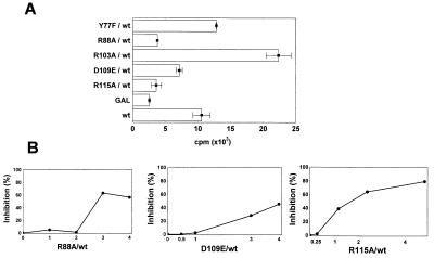
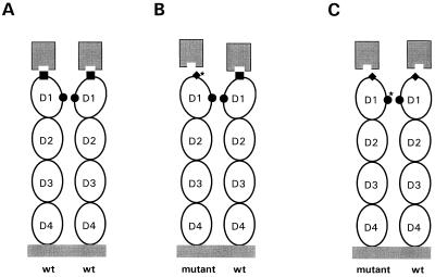
The lymphocyte activation gene-3 (LAG-3), selectively transcribed in human activated T and NK cells, encodes a ligand for major histocompatibility complex (MHC) class II molecules. Like CD4, LAG-3 ectodomain is composed of four Ig-like domains (D1-D4). Nothing is known about the LAG-3 regions or residues required to form a stable MHC class II binding site. In contrast to CD4, soluble LAG-3 molecules stably interact with MHC class II molecules expressed on the cell surface. In addition, the first two N-terminal domains of soluble LAG-3 (D1 and D2) molecules, alone, are capable of binding MHC class II. From a LAG-3 model structure, we designed mutants and tested their ability to bind MHC class II molecules in an intercellular adhesion assay. We found residues on the membrane-distal, CDR1-2-containing top face of D1 that are essential for either binding or repulsing MHC class II proteins. Most of these residues are clustered at the base of a large extra-loop structure that is a hallmark of the LAG-3 D1 Ig-like domain. In addition, as for CD4, oligomerization of LAG-3 on the cell surface may be required to form a stable MHC binding site because mutation of three residues in the ABED beta-strands containing side of D1 results in a dominant negative effect (i.e., binding inhibition of coexpressed wild-type LAG-3).


Figures

Figure 1
Figure 1
Sequence alignment of human and murine LAG-3 and CD4. Identical amino acid residues (at least 3 out of 4) through the LAG-3 and CD4 sequences are shown in bold. Numbers refer to the mature human LAG-3 sequence.
Figure 2
Figure 2
MHC class II binding of selected LAG-3 mutants compared with that of wtLAG-3 expressed at different levels. COS-7 cells were transfected with GAL (30 μg/ml) and 3, 10, 30, and 100 μg/ml of pCDM8-wtLAG-3 DNA, respectively (○). Mutant LAG-3 DNA was used at 30 μg/ml. LAG-3 expression is shown as arbitrary fluorescence units (FU) using 15A9 mAb, and class II binding was determined by cpm counting in a cell–cell adhesion assay using 51Cr-labeled Daudi cells.
Figure 3
Figure 3
Coexpression of R88A, D109, or R115A mutant LAG-3 molecules with wtLAG-3 molecules prevents MHC class II Daudi cells from binding to COS-7 cell transfectants. DNA amounts used for transfection were 10 μg/ml for wtLAG-3 (wt) and the negative control (GAL), and 40 μg/ml for mutants (A). Class II binding was determined by cpm counting in a cell–cell adhesion assay using 51Cr-labeled Daudi cells. B shows the inhibition of Daudi cell binding as a function of the amount of R88A, D109E, or R115A DNA transfected. wtLAG-3 DNA (10 μg/ml) and various amounts of mutant DNA (DNA ratio mutant/wt shown in abscissa) were mixed, and pCDM8 DNA was added to maintain the total amount of transfected DNA at 50 μg/ml.
Figure 4
Figure 4
A hypothetical model of LAG-3-MHC class II interaction. Ovals, D1–D4 of LAG-3 ectodomain; □, active MHC class II binding site (site 1); ♦, inactive MHC class II binding site; •, homotypic interaction binding site (site 2); ★, a mutation with a negative effect on MHC class II binding. The MHC class II heterodimer with the LAG-3 binding site is shown at the top of the figure, facing each LAG-3 molecule. For ease of illustration, molecules are not drawn to scale. (A) A wt–wt LAG-3 homodimer binding two MHC class II heterodimers. (B) A site 1 mutant (Y77F, R103A)–wt LAG-3 homodimer still able to bind MHC. (C) A site 2 mutant (R88A, D109E, R115A)–wt LAG-3 homodimer, with an inappropriate conformational rearrangement in D1, unable to bind MHC.

References

    1. Triebel F, Jitsukawa S, Baixeras E, Roman-Roman S, Genevee C, Viegas-Pequignot E, Hercend T. J Exp Med. 1990;171:1393–1405. - PMC - PubMed
    1. Baixeras E, Huard B, Miossec C, Jitsukawa S, Martin M, Hercend T, Auffray C, Triebel F, Piatier-Tonneau D. J Exp Med. 1992;176:327–337. - PMC - PubMed
    1. Huard B, Prigent P, Pages F, Bruniquel D, Triebel F. Eur J Immunol. 1996;26:1180–1186. - PubMed
    1. Huard B, Prigent P, Tournier M, Bruniquel D, Triebel F. Eur J Immunol. 1995;25:2718–2721. - PubMed
    1. Huard B, Gaulard P, Faure F, Hercend T, Triebel F. Immunogenetics. 1994;39:213–217. - PubMed

Publication types

MeSH terms

Associated data