. 1997 May 27;94(11):5798-803.
doi: 10.1073/pnas.94.11.5798.

Reactivation of silenced, virally transduced genes by inhibitors of histone deacetylase

Affiliations

Reactivation of silenced, virally transduced genes by inhibitors of histone deacetylase

W Y Chen et al. Proc Natl Acad Sci U S A. .

Abstract

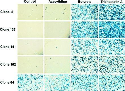
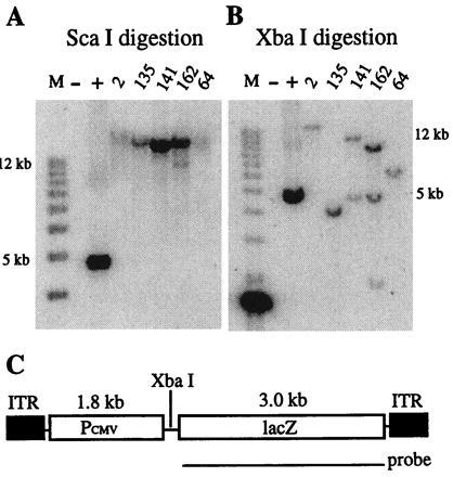
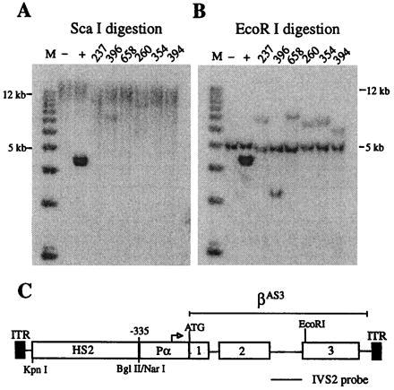
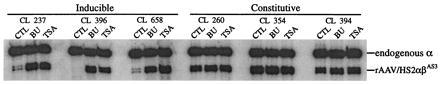
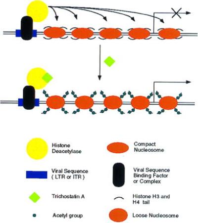
Retroviral and adeno-associated viral sequences can dramatically silence transgene expression in mice. We now report that this repression also occurs in stably infected HeLa cells when the cells are grown without selection. Expression of a transduced lacZ gene (rAAV/CMVlacZ) is silenced in greater than 90% of cells after 60 days in culture. Surprisingly, high-level expression can be reactivated by treating the cells with sodium butyrate or trichostatin A but not with 5-azacytidine. When cell clones with integrated copies of rAAV/CMVlacZ were isolated, lacZ expression was silenced in 80% of the clones; however, lacZ expression was reactivated in all of the silenced clones by treatment with butyrate or trichostatin A. The two drugs also reactivated a silenced globin gene construct (rAAV/HS2alphabetaAS3) in stably infected K562 cells. Trichostatin A is a specific inhibitor of histone deacetylase; therefore, we propose that hyperacetylation of histones after drug treatment changes the structure of chromatin on integrated viral sequences and relieves repression of transduced genes. The reactivation of silenced, transduced genes has implications for gene therapy. Efficient viral gene transfer followed by drug treatment to relieve suppression may provide a powerful combination for treatment of various genetic and infectious diseases.


Figures

Figure 2
Figure 2
X-Gal staining of five representative HeLa cell clones containing integrated rAAV/CMVlacZ. Cells from each clone were assayed for lacZ expression with or without treatment with 5-azacytidine, sodium butyrate, and trichostatin A. Few cells in four of the five untreated controls expressed lacZ; however, both sodium butyrate and trichostatin A dramatically activated lacZ expression. These results are representative of the results obtained with all 25 clones analyzed. One of the 5 clones (clone 64) and 5 of 25 clones overall (data not shown) contained a constitutively active lacZ gene.
Figure 1
Figure 1
Southern blot analysis of HeLa cell clones after rAAV/CMVlacZ infection. HeLa cells were infected with rAAV/CMVlacZ, grown for 3 weeks, and then plated at limiting dilution. Individual cells were expanded for 4 weeks, and DNA was extracted for analysis. Genomic DNA from uninfected HeLa cells was used as negative control, and a 5.0-kb fragment from the rAAV/CMVlacZ vector was spiked into HeLa cell DNA for a positive control. A 3.0-kb lacZ DNA fragment was used as the probe. (A) Genomic DNAs were digested with ScaI, which does not cut rAAV/CMVlacZ. (B) Genomic DNAs were digested with XbaI, which cuts once inside the vector as shown in C. (C) Map of the rAAV/CMVlacZ virus.
Figure 3
Figure 3
Southern blot analysis of rAAV/HS2αβAS3-transduced K562 cell clones. Cells were infected, grown for 30 days, and plated at limiting dilution. Individual cells were expanded for 4 weeks, and DNA was extracted and analyzed as described for HeLa cell clones, except that genomic DNA from uninfected K562 cells was used as a negative control and a 4.5-kb fragment of the rAAV/HS2αβAS3 vector was spiked into K562 cell DNA for a positive control. A 790-bp HinfI–HinfI fragment of the β-globin gene IVS2 was used as a probe. (A) Genomic DNAs were digested with ScaI, which does not cut in rAAV/HS2αβAS3. Hybridization of the probe to high molecular weight bands (greater than 25 kb) demonstrate that viral sequences are not present as episomal vectors. (B) Genomic DNAs were digested with EcoRI, which cuts once in the β-globin gene. The single junction fragments that hybridize to the probe demonstrate that all six clones contain single copy integrants of the transduced gene. The endogenous β-globin gene fragment (5.5 kb) ran slightly faster than expected in this gel. (C) Map of rAAV/HS2αβAS3.
Figure 4
Figure 4
RT-PCR analysis of six representative K562 clones containing integrated rAAV/HS2αβAS3. K562 cell clones were treated with sodium butyrate (BU) or trichostatin A (TSA) for 24 h; untreated cells were included as controls (CTL). Total RNA was extracted with RNA STAT60. RT-PCR of endogenous α-globin mRNA served as an internal control. A dramatic induction of αβAS3 gene expression was observed in half of the clones. The level of butyrate induction was 8.1-fold for clone 237, 33-fold for clone 396, and 9-fold for clone 658. The level of trichostatin A induction was 10.2-fold for clone 237, 19-fold for clone 396, and 14-fold for clone 658. Overall, βAS3 expression was inducible in 6 of the 13 clones, and the average level of induction was 10.6-fold and 9.0-fold for butyrate and trichostatin A, respectively. Expression of αβAS3 in the other half of the samples was constitutive as illustrated by clones 260, 354, and 394.
Figure 5
Figure 5
Model for silencing and reactivation of recombinant viral genes. A host protein or protein complex binds to viral sequences (AAV ITRs or retroviral LTRs) and recruits a histone deacetylase to the site through protein–protein interactions. The enzyme deacetylates histone H3 and H4 N-terminal tails, and the resulting change in chromatin structure inhibits expression from adjacent promoters. Treatment with trichostatin A specifically inhibits histone deacetylases. Subsequent acetylation of histones produces a chromatin structure that allows transcription factors to bind to nearby promoters and activate gene expression.

References

    1. Palmer T D, Rosman G J, Osborne W R A, Miller D A. Proc Natl Acad Sci USA. 1991;88:1330–1334. - PMC - PubMed
    1. Harbers K, Jahner D, Jaenisch R. Nature (London) 1981;293:540–542. - PubMed
    1. Jahner D, Stuhlmann H, Stewart C L, Harbers K, Lohler J, Simon I, Jaenisch R. Nature (London) 1982;298:623–628. - PubMed
    1. McCune S L, Townes T M. Nucleic Acids Res. 1994;22:4477–4481. - PMC - PubMed
    1. Samulski R J, Chang L-S, Shenk T. J Virol. 1989;63:3822–3828. - PMC - PubMed

Publication types

MeSH terms

LinkOut - more resources