. 1997 May 27;94(11):5804-9.
doi: 10.1073/pnas.94.11.5804.

Stable gene transfer and expression of human blood coagulation factor IX after intramuscular injection of recombinant adeno-associated virus

Affiliations

Stable gene transfer and expression of human blood coagulation factor IX after intramuscular injection of recombinant adeno-associated virus

R W Herzog et al. Proc Natl Acad Sci U S A. .

Abstract

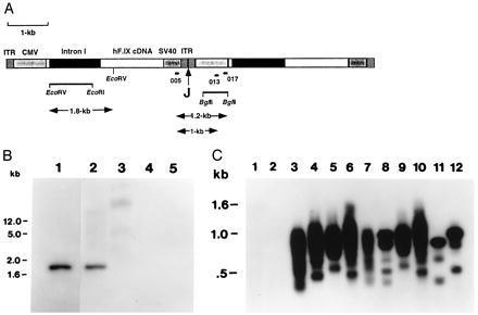
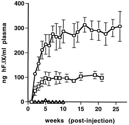
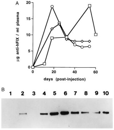
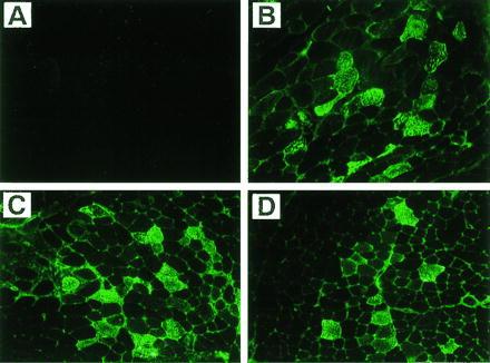
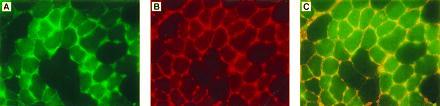
We sought to determine whether intramuscular injection of a recombinant adeno-associated virus (rAAV) vector expressing human factor IX (hF.IX) could direct expression of therapeutic levels of the transgene in experimental animals. High titer (10(12)-10(13) vector genomes/ml) rAAV expressing hF.IX was prepared, purified, and injected into hindlimb muscles of C57BL/6 mice and Rag 1 mice. In the immunocompetent C57BL/6 mice, immunofluorescence staining of muscle harvested 3 months after injection demonstrated the presence of hF.IX protein, and PCR analysis of muscle DNA was positive for AAV DNA, but no hF.IX was detected in mouse plasma. Further studies showed that these mice had developed circulating antibodies to hF.IX. In follow-up experiments in Rag 1 mice, which carry a mutation in the recombinase activating gene-1 and thus lack functional B and T cells, similar results were seen on DNA analysis of muscle, but these mice also demonstrated therapeutic levels (200-350 ng/ml) of F. IX in the plasma. The time course of F.IX expression demonstrates that levels gradually increase over a period of several weeks before reaching a plateau that is stable 6 months after injection. In other experiments we demonstrate colocalization of hF.IX and collagen IV in intersitial spaces between muscle fibers. Collagen IV has recently been identified as a F.IX-binding protein; this finding explains the unusual pattern of immunofluorescent staining for F.IX shown in these experiments. Thus rAAV can be used to direct stable expression of therapeutic levels of F.IX after intramuscular injection and is a feasible strategy for treatment of patients with hemophilia B.


Figures

Figure 5
Figure 5
Analyses of DNA isolated from muscle injected with AAV-F.IX. (A) Diagram showing head-to-tail tandem repeats of two vector genomes of AAV-F.IX. Indicated are AAV inverted terminal repeats (ITR), CMV promoter/enhancer (CMV), hF.IX cDNA including coding sequence, and 228 bp of the 3′ untranslated region, a 1.4-kb portion of intron I, simian virus 40 polyadenylylation signal (SV40), and the junction site of the two genomes (J). A 1.2-kb EcoRV–EcoRI fragment from intron I and a 0.7-kb BglII fragment from the CMV promoter fragment were chosen as probes for Southern blot hybridization. Positions of binding sites for primers 005 (forward primer), 013, and 017 (reverse primers) are also shown. (B) Southern blot hybridization of genomic DNA isolated from muscle of Rag 1 mouse 6 weeks postinjection with AAV-F.IX. A radioactively labeled EcoRV–EcoRI fragment from intron I of hF.IX served as a probe. Lane 1, pAAV-FIX plasmid DNA (50 pg). Lanes 2 and 3, DNA isolated from muscle injected with AAV-F.IX. Lanes 4 and 5, DNA isolated from uninjected animal. Lanes 1, 2, and 4, DNA digested with EcoRV. Lanes 3 and 5, undigested DNA. Fifteen micrograms of genomic DNA per lane (lanes 2–5). DNA was separated on a 1% agarose gel before transfer onto a nylon membrane (Schleicher and Schuell). Size markers in the left margin are based on migration of a commercially available set of markers (GIBCO/BRL). (C) Southern blot hybridization of junction fragments of head-to-tail concatemers of AAV-F.IX amplified by PCR. PCR products amplified from genomic DNA using primer pair 005–013 (odd-numbered lanes) or primer pair 005–017 (even-numbered lanes) are shown. Lanes 1 and 2, Uninjected animal. Lanes 3–6, C57BL/6 mice i.m.-injected with AAV-F.IX. Lanes 7–10, Rag 1 mice i.m.-injected with AAV-F.IX. PCR products were from tibialis anterior (lanes 3, 4, 7, and 8) or quadriceps (lanes 5, 6, 9, and 10) muscle DNA. Lanes 11 and 12, PCR products from cell line 10–3.AV 5, which contains at least two monomer copies of integrated AAV-lacZ arranged head-to-tail (10). Note that the sizes of the major PCR products for lanes 11 and 12 are 0.8 and 1 kb, respectively. PCR products were separated on a 2% agarose gel before blotting onto a nylon membrane. A 0.7-kb BglII fragment from the CMV promoter served as a probe. Genomic muscle DNA had been isolated 6 to 8 weeks postinjection.
Figure 1
Figure 1
Plasma concentration of hF.IX in experimental mice as a function of time after intramuscular injection with AAV-hF.IX. ▵, C57BL/6 mice after i.m. injection of 2 × 10¹¹ vector genomes/animal. □, Rag 1 mice after i.m. injection of 1 × 10¹⁰ vector genomes/animal. ○, Rag 1 mice after i.m. injection of 2 × 10¹¹ vector genomes/animal. Each line represents an average of four animals with error bars denoting SD.
Figure 2
Figure 2
Circulating antibody against hF.IX as a result of i.m. injection of AAV-F.IX in C57BL/6 mice. (A) Time course of anti-hF.IX antibody concentration in plasma after injection with 2 × 10¹¹ vector genomes/animal (n = 3) as determined by ELISA using mouse monoclonal anti-hF.IX (Boehringer Mannheim) as a standard. Each line represents an individual animal. Note that the units on the y axis represent an estimate only, because of probable differences in affinity for hF.IX between the mouse monoclonal used as the standard and the antibodies present in the serum of injected mice. (B) Western blot demonstrating the presence of antibodies against hF.IX in plasma of C57BL/6 mice after i.m. injection. Lane 1, Animal injected i.m. with AAV-lacZ, day 18 postinjection. Lane 2, Animal injected i.m. with rAAV-hF.IX (14), day 20 postinjection. Lanes 3–10, Animals injected i.m. with AAV-F.IX. Lanes 3–7, days 11, 18, 32, 54, and 60 postinjection (same animal). Lanes 8–10, day 18 postinjection (different animals).
Figure 3
Figure 3
Immunofluorescence staining (with antibody to hF.IX) of tibialis anterior muscle of C57BL/6 mice. (A) Uninjected muscle. (B–D) Muscle 3 months postinjection with AAV-F.IX (3.3 × 10¹⁰ vector genomes per injection site). (×200.)
Figure 4
Figure 4
Immunofluorescence staining of muscle sections of tibialia anterior muscle of C57BL/6 mice injected with AAV-F.IX 3 months earlier. Muscle sections were stained simultaneously with FITC-conjugated antibody against hF.IX and a rhodamine-conjugated antibody complex against collagen IV. (A) Fluorescence of FITC (green) showing the presence of hF.IX in muscle fibers and interstitial spaces. (B) Fluorescence of rhodamine (red) showing collagen IV in the extracellular matrix of muscle fibers. (C) Simultaneous excitation of both fluorescence tags. Note the presence of a yellow signal in the interstitial spaces. (×400.)

References

    1. Dai Y, Schwartz E M, Gu D, Zhang W W, Sarvetnick N, Verma I M. Proc Natl Acad Sci USA. 1995;92:1401–1405. - PMC - PubMed
    1. Tripathy S K, Black H B, Goldwasser E, Leiden J M. Nat Med. 1996;2:545–550. - PubMed
    1. Yang Y, Haecker S E, Su Q, Wilson J M. Hum Mol Genet. 1996;5:1703–1712. - PubMed
    1. Tripathy S K, Svensson E C, Black H G, Goldwasser E, Margalith M, Hobart P M, Leiden J M. Proc Natl Acad Sci USA. 1996;93:10876–10880. - PMC - PubMed
    1. Carter B J. Curr Opin Biotech. 1992;3:533–539. - PubMed

Publication types

MeSH terms