. 1997 May 27;94(11):5843-7.
doi: 10.1073/pnas.94.11.5843.

Apoptin induces apoptosis in human transformed and malignant cells but not in normal cells

Affiliations

Apoptin induces apoptosis in human transformed and malignant cells but not in normal cells

A A Danen-Van Oorschot et al. Proc Natl Acad Sci U S A. .

Abstract

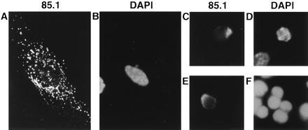
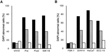
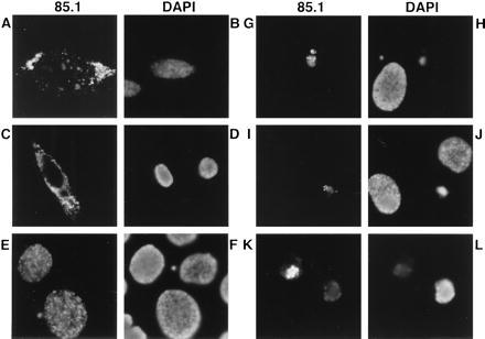
The chicken anemia virus protein apoptin induces a p53-independent, Bcl-2-insensitive type of apoptosis in various human tumor cells. Here, we show that, in vitro, apoptin fails to induce programmed cell death in normal lymphoid, dermal, epidermal, endothelial, and smooth-muscle cells. However, when normal cells are transformed they become susceptible to apoptosis by apoptin. Long-term expression of apoptin in normal human fibroblasts revealed that apoptin has no toxic or transforming activity in these cells. In normal cells, apoptin was found predominantly in the cytoplasm, whereas in transformed and malignant cells it was located in the nucleus, suggesting that the localization of apoptin is related to its activity. These properties make apoptin a potential agent for the treatment of a large number of tumors, also those lacking p53 and/or overexpressing Bcl-2.


Figures

Figure 1
Figure 1
Localization of apoptin and staining of the chromatin in three normal human cell types, transiently transfected with pCMV-fs. Apoptin was stained with anti-apoptin mAb 85.1 (A, C, and E) and the DNA was stained with DAPI (B and D) or PI (F), in representations of identical cells: (A and B) HSMC, (C and D) HUVEC, and (E and F) T cells. Cells were fixed 5 days after transfection and analyzed by indirect immunofluorescence. (Original magnification: A and B, ×630; C–F, ×1,000.)
Figure 2
Figure 2
Apoptin activity in normal versus transformed/malignant cells. The percentage of cells that stained abnormally with DAPI is given as a relative measure for apoptosis in (A) normal (VH10) versus transformed (pre, post) and tumor (NW-18) fibroblasts and (B) normal (FSK-1) versus transformed (SVK14, HaCaT) and tumor (SCC-15) keratinocytes transiently transfected with pCMV-fs (solid bars), pCMV-tr (shaded bars), or pCMV-des (open bars). Cells were fixed 5 days after transfection and analyzed by indirect immunofluorescence. Results are the means of at least three independent experiments. In each experiment at least 200 cells were examined that were positive for full-sized or truncated apoptin, or for desmin.
Figure 3
Figure 3
Localization of apoptin and staining of the chromatin in normal and transformed/malignant cells transiently transfected with pCMV-fs. Apoptin was stained with anti-apoptin mAb 85.1 and DNA with DAPI in representations of identical cells: (A and B) VH10, (C and D) FSK-1, (E and F) HaCaT, (G and H) post, (I and J) NW-18, and (K and L) SCC-15. Cells were fixed 2 (E and F) or 5 (A–D and G–L) days after transfection and analyzed by indirect immunofluorescence. (Original magnification: A–D, ×630; I–L, ×1,000; and E–H, ×1,250.)

Similar articles

Cited by

References

    1. Earnshaw W C. Curr Opin Cell Biol. 1995;7:337–343. - PubMed
    1. Wyllie A H, Kerr J F R, Currie A R. Int Rev Cytol. 1980;68:251–306. - PubMed
    1. Arends M J, Wyllie A H. Int Rev Exp Pathol. 1991;32:223–254. - PubMed
    1. Roy C. Exp Cell Res. 1992;200:416–424. - PubMed
    1. Wyllie A H. Curr Opin Genet Dev. 1995;5:97–104. - PubMed

Publication types

MeSH terms

Substances