. 1997 May 27;94(11):5906-11.
doi: 10.1073/pnas.94.11.5906.

The neuron-restrictive silencer element: a dual enhancer/silencer crucial for patterned expression of a nicotinic receptor gene in the brain

Affiliations

The neuron-restrictive silencer element: a dual enhancer/silencer crucial for patterned expression of a nicotinic receptor gene in the brain

A Bessis et al. Proc Natl Acad Sci U S A. .

Abstract

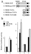
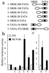
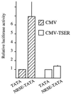
The neuron-restrictive silencer element (NRSE) has been identified in several neuronal genes and confers neuron specificity by silencing transcription in nonneuronal cells. NRSE is present in the promoter of the neuronal nicotinic acetylcholine receptor beta2-subunit gene that determines its neuron-specific expression in the nervous system. Using transgenic mice, we show that NRSE may either silence or enhance transcription depending on the cellular context within the nervous system. In vitro in neuronal cells, NRSE activates transcription of synthetic promoters when located downstream in the 5' untranslated region, or at less than 50 bp upstream from the TATA box, but switches to a silencer when located further upstream. In contrast, in nonneuronal cells NRSE always functions as a silencer. Antisense RNA inhibition shows that the NRSE-binding protein REST contributes to the activation of transcription in neuronal cells.


Figures

Figure 1
Figure 1
Point mutation of NRSE in the promoter of the nAChR β2-subunit gene changes the pattern of β-gal expression in transgenic mice. (ad) Whole-mount coloration of E13.5 transgenic embryos carrying the wild-type promoter (a and c) and with point mutation in NRSE (b and d). (e and f) Detection of the β-gal activity and the glial fibrillary acidic protein immunoreactivity in the adult mutated transgenic brain. (e) Dorsal part of the hippocampus. (f) Dorso-medial part of the cortex, showing the absence of β-gal expression in the molecular layer (arrow). og, orthosympathetic ganglia; drg, dorsal root ganglia; Co, colliculus; Th, thalamus; Cx, cortex; Hip, hippocampus; cc, corpus callosum.
Figure 2
Figure 2
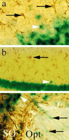
Expression of the NRSE-mutated nAChR β2-subunit gene promoter is restricted to neurons. The β-gal activity (arrowheads) and the glial fibrillary acidic protein immunoreactivity (arrows) were compared in different regions of the adult brain. (a) Layer 2 of the pyriform cortex. (b) Pyramidal neurons of the hippocampus. (c) Oligodendrocytes (arrows) of the optic tract (Opt) and neurons (arrowheads) of the Supra-optic nucleus (SO).
Figure 3
Figure 3
Enhancer and silencer activities of NRSE within synthetic promoters. (a) Schematic representation of the plasmids used for transfection. Oval, three NRSEs; crossed-out oval, mutated NRSEs; solid box, SV40 TATA box; hatched box, myosin IIB TATA box. (b) Relative activity of these plasmids transfected in different cell lines. The luciferase activities were normalized to that of the same plasmid containing no NRSE sequences (SVP-Luci, TATA-Luci, or TATAIIB-Luci).
Figure 4
Figure 4
The sign of the regulatory activity of NRSE depends on the primary structure of the promoter in neuroblastomas but not in fibroblasts. (a) Schematic representation of the plasmids. See Fig. 3 legend for explanations of symbols. (b and c) Relative activities of the plasmids transiently transfected. The activities of the plasmids were normalized to that of the same plasmids with no NRSE.
Figure 5
Figure 5
Detection of NRSE binding activity and REST mRNA in fibroblasts and neuroblastoma. (Left) Mobility shift experiment using an NRSE probe and extracts from fibroblast (lanes 1–4) or neuroblastoma cells (lanes 5–8). The gel retardation was done in presence of 100-fold molar excess of cold NRSE (lanes 2 and 6), NRSEmut (lanes 3 and 7), or oligonucleotide SP1 carrying an SP1 site (lanes 4 and 8). (Right) RT-PCR with RNA extracted from fibroblasts (lanes 1 and 2, one round of PCR amplification) or neuroblastomas (lanes 3 and 4, two rounds of PCR amplification using nested primers). Lanes 2 and 4, reverse transcriptase was omitted.
Figure 6
Figure 6
REST trans-activates transcription through binding to NRSE. Activation of transcription by NRSE was compared after cotransfection with the CMV-TSER plasmid expressing a REST antisense mRNA or with the expression plasmids carrying no cDNA. The activity of the NRSE-TATA plasmid is compared with that of TATA-plasmid cotransfected with the same expression plasmid set to 1.

References

    1. Johnson J E, Birren S J, Anderson D J. Nature (London) 1990;346:858–861. - PubMed
    1. Akazawa C, Sasai Y, Nakanishi S, Kageyama R. J Biol Chem. 1992;267:21879–21885. - PubMed
    1. Lee J, Hollenberg S, Snide R L, Turner D, Lipnick N, Weintraub H. Science. 1995;268:836–844. - PubMed
    1. Bessis A, Savatier N, Devillers-Thiery A, Bejanin S, Changeux J P. Nucleic Acids Res. 1993;21:2185–2192. - PMC - PubMed
    1. Bessis A, Salmon A, Zoli M, Le Novere N, Picciotto M, Changeux J P. Neuroscience. 1995;69:807–819. - PubMed

Publication types

MeSH terms

LinkOut - more resources