Review
. 1997 Mar 1;7(3):R186-95.
doi: 10.1016/s0960-9822(97)70085-x.

Limb morphogenesis: connections between patterning and growth

Affiliations
Review

Limb morphogenesis: connections between patterning and growth

N Serrano et al. Curr Biol. .

Abstract

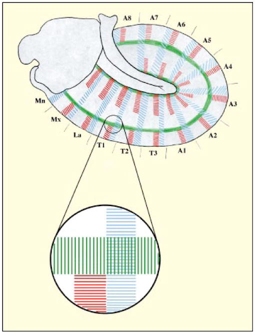
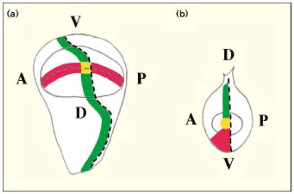
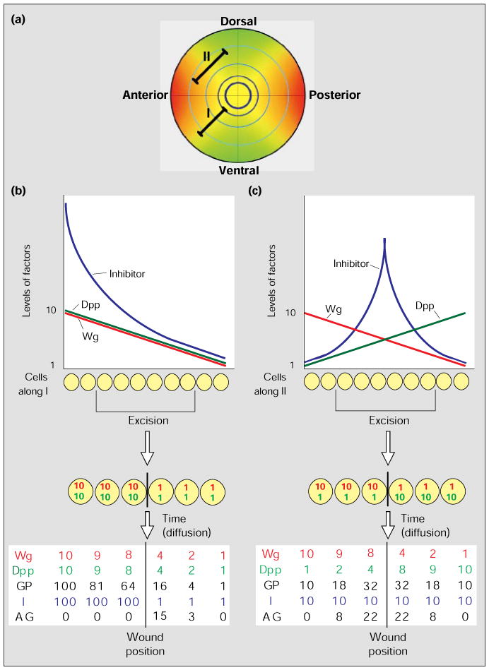
Limb development is a complex process involving precise control of both patterning and growth. Great strides have been made in understanding limb morphogenesis and identifying essential patterning genes in Drosophila. Differential expression of these genes divides the future limb into territories, which will give rise to different regions of the adult appendage. Recent analyses have defined the role of territorial boundaries as organizers of both patterning and growth, highlighting the connection between these two processes. The organizing activity of territorial boundaries seems to be mediated through the activity of two locally produced morphogens: Wingless and Decapentaplegic. We propose a model in which these two molecules, distributed in a graded fashion, act in synergy to promote growth of the entire appendage. We also suggest that existence of growth inhibitors that counteract the action of Wingless and Decapentaplegic; by opposing the gradient of these growth factors, the inhibitors guide the near-uniform proliferation that shapes the imaginal discs from which the adult appendages are formed in Drosophila.


Figures

Figure 1
Figure 1
The intersection of En, Dpp and Wg expression stripes defines the position of imaginal disc primordia in Drosophila. The main diagram is a lateral view of a stage 11 Drosophila embryo. Engrailed (En) expression in the posterior compartment cells is shown in blue, Wingless (Wg) expression in adjacent anterior compartment cells in red and Decapentaplegic (Dpp) in green. The circle represents the region where the first leg disc primordium forms, and an enlarged view of this is also shown. Segment abbreviations: Mn, mandibulary; Mx, maxillary; La, labial; T1–3, thoracic segments; A1–8, abdominal segments.
Figure 2
Figure 2
A duplicated Drosophila wing generated by a posterior clone of en/inv mutant cells [12]. The veins in the original wing are indicated by numbers, and the veins in the duplicated wing by primed numbers. The arrow indicates the position of the clone.
Figure 3
Figure 3
A diagram showing how expression stripes of development genes cross in the Drosophila wing disc (a) but meet in the leg disc (b). The dashed lines represent the boundaries between the anterior (A) and posterior (P), and the dorsal (D) and ventral (V), compartments. Red represents Wg expression, and green Dpp expression; yellow dots represent the regions where Wg and Dpp expression domains intersect, or meet, at the center of the imaginal discs. This central region will give rise to the distal-most structures of the appendages.
Figure 4
Figure 4
The Drosophila wing pattern is reorganized as a result of ectopic Dpp expression [25]. The generation of a Dpp-expressing clone in the anterior compartment of the wing disc, which contributes to both the dorsal and the ventral surfaces of the wing, induces formation of an ectopic, double-anterior wing. The clone borders are outlined in blue ventrally and in red dorsally; the numbers indicate the veins.
Figure 5
Figure 5
A model for intercalary growth in Drosophila imaginal discs. (a) An idealized diagram of the graded distribution of Wg and Dpp within the wing disc. The distributions of Wg (red) and Dpp (green) expression within the disc are shown by the gradients of color intensity; yellow represents regions where Wg and Dpp expression overlap. The intersecting lines represent the anterior–posterior (vertical line) and dorsal–ventral (horizontal line) boundaries. The pale blue concentric circles are meant to represent proximal–distal distinctions that might result from the joint action of high levels of Dpp and Wg at successive times during the growth of the disc. Lines I and II represent rows of cells: the cells along I maintain the same ratio of Dpp and Wg levels – see (b) – but the absolute levels drop with distance from the sources of morphogen; cells along II differ dramatically in the ratio of Dpp and Wg levels – see (c) – depending on the relative distance from the Wg and Dpp sources. (b,c) The graphs show how the concentration levels, in arbitrary numbers, of Wg (red), Dpp (green) and a hypothetical growth inhibitor (blue) vary along lines I (b) and II (c); we have used linear gradients of morphogen concentration for simplicity, but they could well be exponential. Below the graphs we illustrate the consequence of an excision of some of the cells, which juxtaposes cells from distant positions giving discontinuities in the gradients of Wg (red) and Dpp (green). The table of numbers at the bottom gives the expected levels of the growth factors Wg and Dpp after some diffusion across the wound. Using the model, the table also indicates the growth potential (GP) based on synergy between Dpp and Wg (GP = W × D), the presumed level of inhibitor (I) and the net or actual growth (AG). All numbers are arbitrary and are assigned only to illustrate the principle.

References

    1. Spemann H, Mangold H. Uber induktion von embryonenanlagen durch implantation artfremder organisatoren. Arch Mikrosk Anat Entw Mech. 1924;100:599–638.
    1. Tickle C, Summerbell D, Wolpert L. Positional signalling and specification of digits in chick limb morphogenesis. Nature. 1975;254:199–202. - PubMed
    1. Cohen SM. Imaginal disc development. In: Bate M, Martinez-Arias A, editors. The Development of Drosophila melanogaster. Plainview, New York: Cold Spring Harbor Laboratory Press; 1993. pp. 747–841.
    1. Cohen B, Simcox AA, Cohen SM. Allocation of the thoracic imaginal primordia in the Drosophila embryo. Development. 1993;117:597–608. - PubMed
    1. Garcia-Bellido A. Cell Patterning CIBA symposium. Boston: Little Brown; 1975. Genetic control of wing disc development in Drosophila; pp. 161–183. - PubMed

MeSH terms

LinkOut - more resources