. 1997 Mar;4(3):231-8.
doi: 10.1038/nsb0397-231.

Crystal structures of HINT demonstrate that histidine triad proteins are GalT-related nucleotide-binding proteins

Affiliations

Crystal structures of HINT demonstrate that histidine triad proteins are GalT-related nucleotide-binding proteins

C Brenner et al. Nat Struct Biol. 1997 Mar.

Abstract

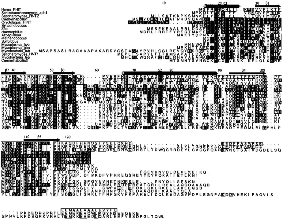
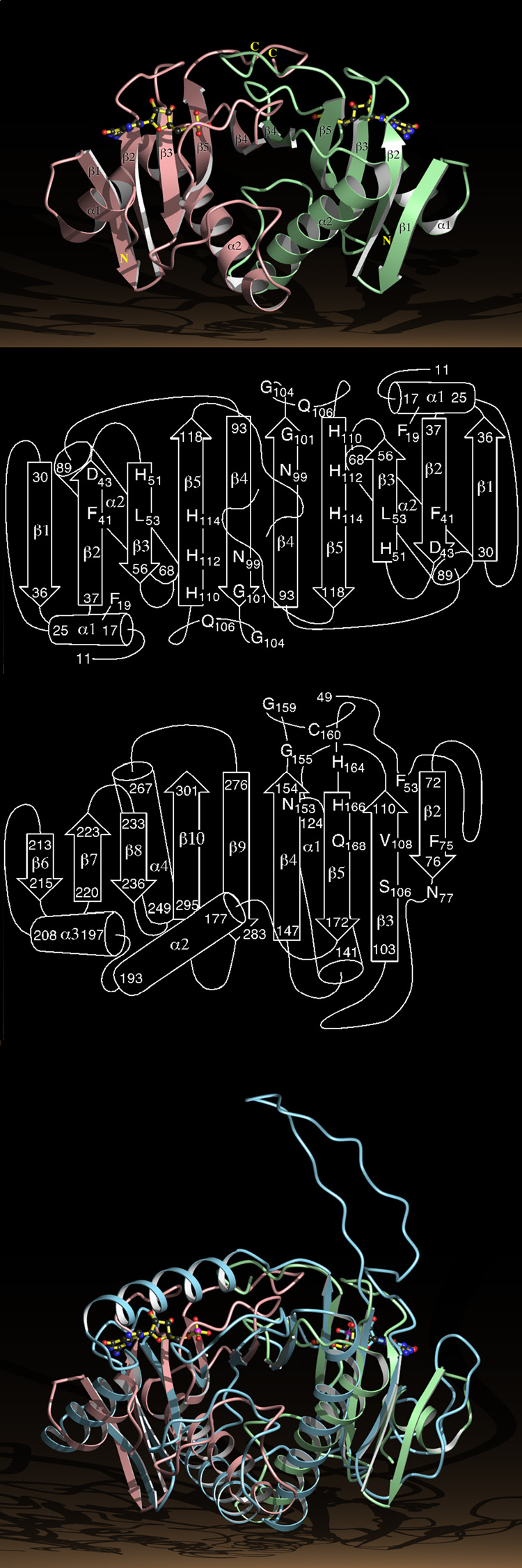
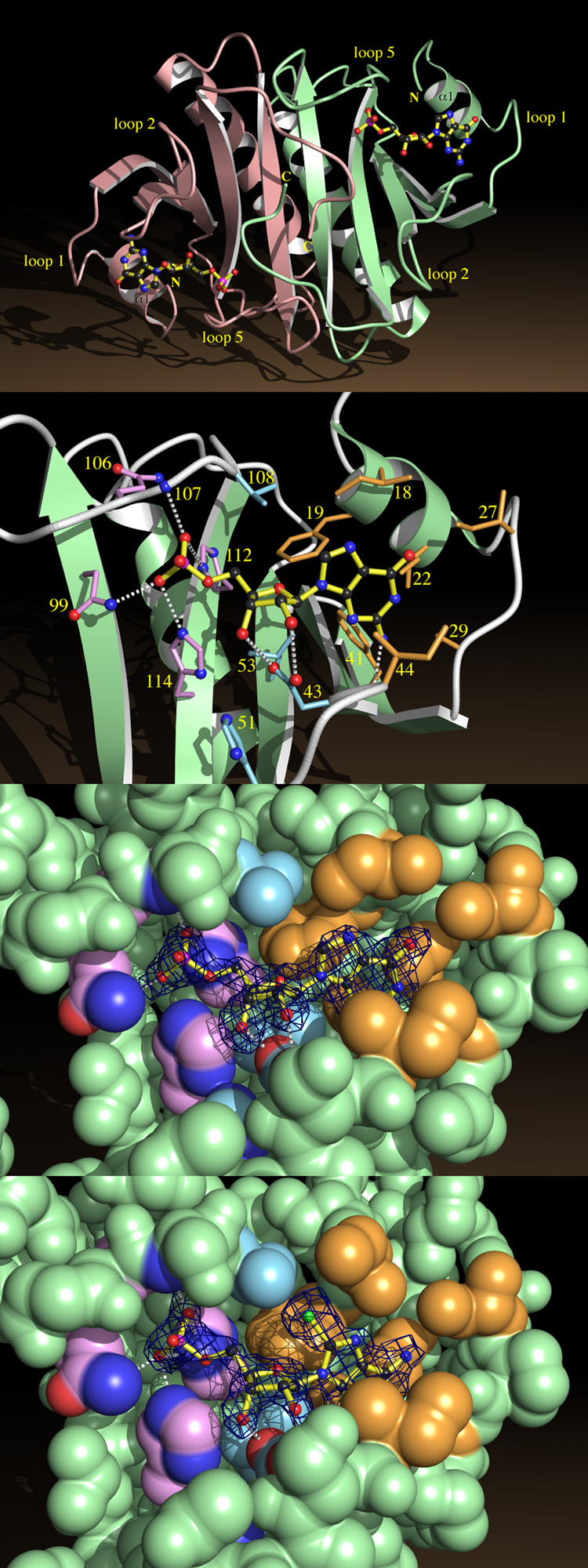
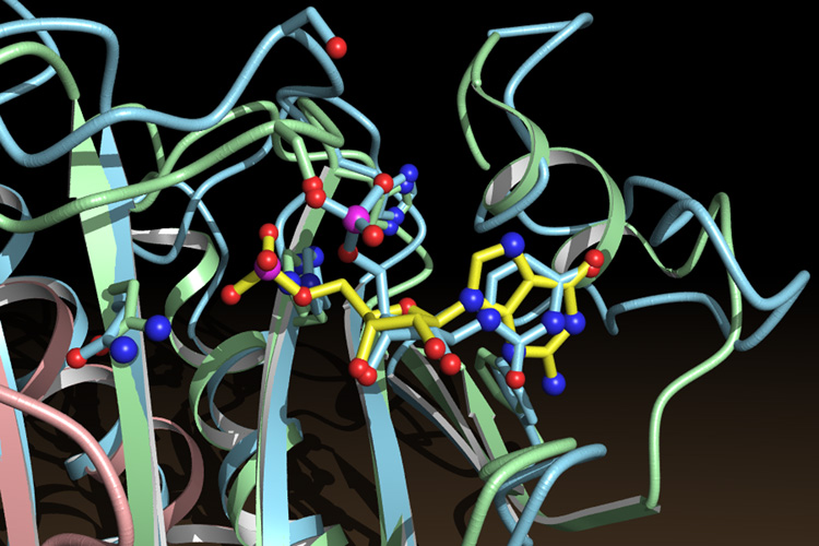
Histidine triad nucleotide-binding protein (HINT), a dimeric purine nucleotide-binding protein from rabbit heart, is a member of the HIT (histidine triad) superfamily which includes HINT homologues and FHIT (HIT protein encoded at the chromosome 3 fragile site) homologues. Crystal structures of HINT-nucleotide complexes demonstrate that the most conserved residues in the superfamily mediate nucleotide binding and that the HIT motif forms part of the phosphate binding loop. Galactose-1-phosphate uridylyltransferase, whose deficiency causes galactosemia, contains tandem HINT domains with the same fold and mode of nucleotide binding as HINT despite having no overall sequence similarity. Features of FHIT, a diadenosine polyphosphate hydrolase and candidate tumour suppressor, are predicted from HINT-nucleotide structures.


Figures

Fig. 1
Fig. 1
Sequence alignment of HIT proteins excluding nearly identical proteins, expressed sequence tags and partial sequences. Strict conservation of residues aligning with Phe 19, His 51, Leu 53, and Histidines 110, 112 and 114 was required for inclusion. The top three proteins are members of the FHIT branch. The others are members of the HINT branch. Genera refer their most common species, except Mycobacteria_tub which refers to M. tuberculosis; Mycoplasma_gen, M. genetalium; Mycoplasma_hyo, M. hyorhinis; Mycobacteria_lep, M. leprae. Black boxes designate identity with rabbit (Oryctologus) HINT, white boxes identity with human FHIT at a residue not conserved with HINT. Secondary structure assignments and numbering are those of HINT.
Fig. 2
Fig. 2
Gel Filtration Chromatography of HINT. A Sephadex G-100 column was calibrated with carbonic anhydrase (MW = 29 kDa) and lysozyme (MW = 14.3 kDa) before HINT was analyzed. 0.5 ml fractions were collected. Peaks were detected by optical density.
Fig. 3
Fig. 3
Structure of the HINT Dimer and the GalT Monomer. a, Ribbon diagram of the HINT-GMP dimer with numbered secondary structural elements. The right hand HINT monomer is in green, the left in pink. b, Secondary structure of the HINT dimer with the positions of a subset of the conserved residues in the HIT superfamily. c, Secondary structure of residues 49 to 302 of the GalT core identifying residues that align in three dimensions with HINT. d, The Cα positions of residues 49 to 302 of the GalT core, represented by a ribbon diagram in blue, superimposed on the HINT-GMP dimer.
Fig. 4
Fig. 4
Nucleotide Binding by HINT. a, HINT-GMP dimer shown from above the β-sheet, indicating helices α1 and loops 1, 2 and 5. b, A close-up of GMP recognition by HINT including nonhydrogen atoms of GMP and side chains from fifteen conserved residues. Side chains surrounding the guanine base are beige, those surrounding ribose are blue, and those surrounding phosphate are purple. Potential hydrogen bonds between GMP and protein are indicated. c, Coordinates corresponding to GMP in the HINT-GMP structure were removed and the protein model was subjected to simulated annealing at 3000 °C. A difference electron density map was calculated with coefficients (Fobs - Fcalc) and contoured at 2.4 σ over the omitted GMP coordinates. Agreement between the map and model of GMP demonstrates that phases calculated from the protein model are robust and that GMP binds in this manner. The nucleotide-binding site is represented by its van der Waals surface, color coded as above. d, A simulated annealing omit map prepared, as above, for HINT-8-Br-AMP.
Fig. 5
Fig. 5
Superposition of the nucleotide-binding sites of HINT and GalT. GMP (yellow bonds) belongs to the HINT protein (green and pink). UMP (blue bonds) belongs to the GalT protein (blue).

References

    1. Seraphin B. The HIT protein family: a new family of proteins present in prokaryotes yeast and mammals. DNA Sequence. 1992;3:177–179. - PubMed
    1. Mozier NM, Walsh MP, Pearson JD. Characterization of a novel zinc binding site of protein kinase C inhbitor-1. FEBS Lett. 1991;279:14–18. - PubMed
    1. McDonald JR, Walsh MP. Ca2+-binding proteins from bovine brain including a potent inhibitor of protein kinase C. Biochem. J. 1985;232:559–567. - PMC - PubMed
    1. Brzoska PM, et al. The product of the ataxia-telangiectasia group d complementing gene, atdc interacts with a protein kinase c substrate and inhibitor. Proc. Natl. Acad. Sci. USA. 1995;92:7824–7828. - PMC - PubMed
    1. Lima C, Klein MG, Weinstein IB, Hendrickson WA. Three-dimensional structure of human protein kinase C interacting protein 1, a member of the HIT family of proteins. Proc. Natl. Acad. Sci. USA. 1996;93:5357–5362. - PMC - PubMed

Publication types

MeSH terms

LinkOut - more resources