Review
. 1996;74(6):833-51.
doi: 10.1139/o96-089.

The importance of the microenvironment in breast cancer progression: recapitulation of mammary tumorigenesis using a unique human mammary epithelial cell model and a three-dimensional culture assay

Affiliations
Review

The importance of the microenvironment in breast cancer progression: recapitulation of mammary tumorigenesis using a unique human mammary epithelial cell model and a three-dimensional culture assay

V M Weaver et al. Biochem Cell Biol. 1996.

Abstract

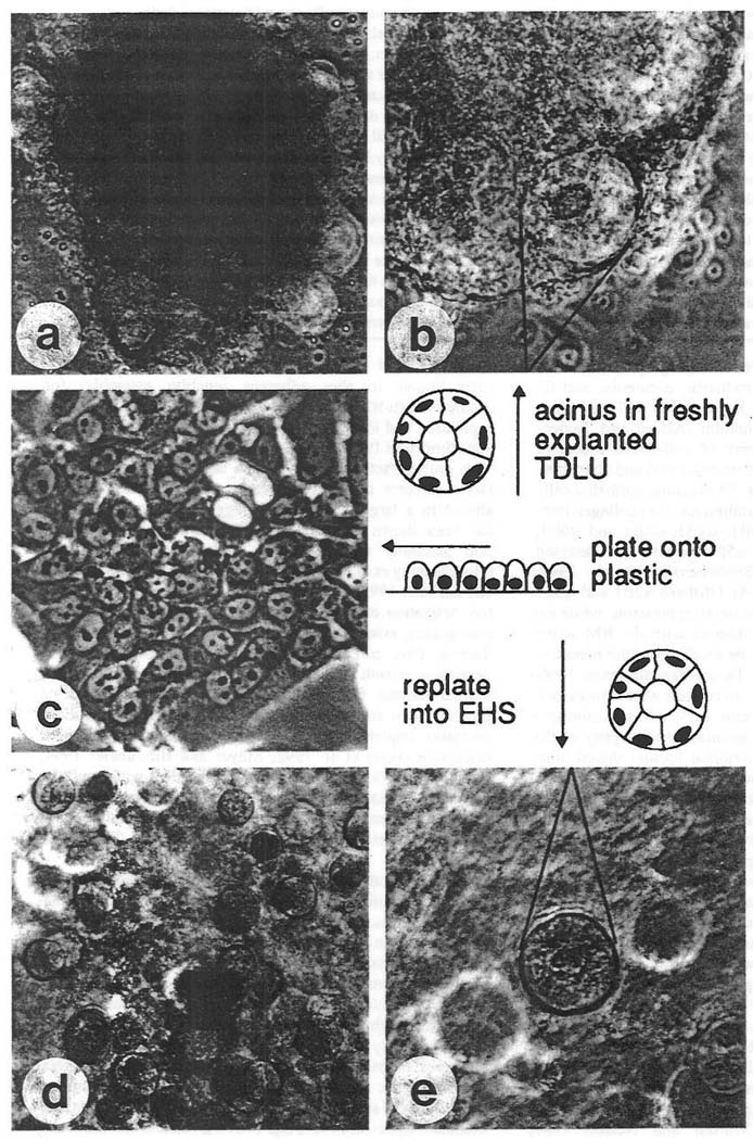
The extracellular matrix (ECM) is a dominant regulator of tissue development and homeostasis. "Designer microenvironments" in culture and in vivo model systems have shown that the ECM regulates growth, differentiation, and apoptosis in murine and human mammary epithelial cells (MEC) through a hierarchy of transcriptional events involving the intricate interplay between soluble and physical signaling pathways. Furthermore, these studies have shown that these pathways direct and in turn are influenced by the tissue structure. Tissue structure is directed by the cooperative interactions of the cell-cell and cell-ECM pathways and can be modified by stromal factors. Not surprisingly then, loss of tissue structure and alterations in ECM components are associated with the appearance and dissemination of breast tumors, and malignancy is associated with perturbations in cell adhesion, changes in adhesion molecules, and a stromal reaction. Several lines of evidence now support the contention that the pathogenesis of breast cancer is determined (at least in part) by the dynamic interplay between the ductal epithelial cells, the microenvironment, and the tissue structure (acini). Thus, to understand the mechanisms involved in carcinogenesis, the role of the microenvironment (ECM as well as the stromal cells) with respect to tissue structure should be considered and studied. Towards this goal, we have established a unique human MEC model of tumorigenesis, which in concert with a three-dimensional assay, recapitulates many of the genetic and morphological changes observed in breast in cancer in vivo. We are currently using this system to understand the role of the microenvironment and tissue structure in breast cancer progression.


Figures

Fig. 1
Fig. 1
Tissue structure, as determined by the dynamic interactions between the cell and the extracellular matrix (ECM), via its cell adhesion molecules (CAMs), as an integrator of function in the mammary gland. The ECM by its effects on tissue structure and through the CAMs, positively influences tissue-specific gene expression and inhibits inappropriate branching, proliferation, apoptosis, and the development of cancer.
Fig. 2
Fig. 2
Phase-contrast micrographs of normal primary breast epithelial cells. (a) Freshly explanted terminal duct lobular unit at low magnification (×100). (b) Single acinus in focus at a higher magnification (×250). (c) Second passage cells in monolayer culture (×250). (d) Fully developed spheres in the EHS matrix (×100). (e) Single sphere at higher magnification, (x250). (Reproduced with permission from Petersen et al. 1992.)
Fig. 3
Fig. 3
Photo-contrast micrographs of tissue sections stained with hematoxylin and eosin of normal human breast tissue (a), ductal epithelial hyperplasia (b), atypical ductal hyperplasia (c), ductal carcinoma in situ (DCIS) (d), DCIS with invasive ductal carcinoma (e), and a fine needle aspirate of a ductal carcinoma (f). (a) In the normal breast, multiple cell types contribute to the organization of the ducts. On the innermost part of the duct (small arrows) are the ductal epithelial cells. External to the ductal cells are myoepithelial cells (arrowheads) derived from terminal differentiation of the ductal cells. The abundant collagen and other extracellular matrix material of the stroma (appearing fibrillar) wraps concentrically around the ducts. The stroma is arranged concentrically around the entire lobule (delineated by long arrows) and then around each individual acinus (×200). (b) In ductal epithelial hyperplasia, the ductal cells have grown towards the center of the duct and formed small bridges or arches across the lumen of the duct. The ductal cells that grow in their native environment next to the basal lamina (long thin arrows) have a more active nuclear appearance than the cells farthest from the basal lamina (long thick arrows). The activity of the nucleus is gauged on the basis of the degree of heterochromatin formation. The presence of abundant heterochromatin in the central-most ductal cells predicts that gene transcription has decreased in these cells compared with the cells toward the basal lamina zone (×400). Residual myoepithelial cells (small arrows) are still present. (c) In atypical ductal hyperplasia, the ductal cells are able to grow away from the basal lamina microenvironment (delineated by long, thin arrows) with almost no effect on their degree of activation. Furthermore, the myoepithelial cells become sparse. Note the presence of some cells with slightly more heterochromatin and less cytoplasm than the rest of the cells (compare curved arrow with open arrow cells). Also, the ductal cells focally have a shared polarity in a few areas of the duct (see the lower leftmost bridge of cells just below the curved arrow) (×200). (d) The defining feature of ductal carcinoma is the apparent complete trophic independence of the ductal cells from the basal lamina zone (delineated by short arrows). Thus, the ductal cells can grow away from the basal lamina zone into the center of the duct (arrow) and maintain identical nuclear and cytoplasmic features compared with the cells next to the native basal lamina zone (×400). (e) On the right is a focus of DCIS in which the original concentric wrapping of stromal collagen is intact, the outline of the duct is smooth, and the fibroblasts next to the DCIS (curved arrow) appear inactive since the nucleus is small and heterochromatic. In contrast, ragged groups of infiltrating ductal cells without the normal organized wrapping of collagen is seen on the left side. The invasive cells appear able to activate the stromal fibroblasts since the fibroblasts next to the invasive ductal cells show euchromatic nuclei (straight arrow) with occasional nucleoli. This diagnostic apparent tropic interaction between infiltrating cancer cells and the stroma is called desmoplasia (×400). (f) Ductal cells strip away from the stroma in fine needle aspirates and invasion is difficult to diagnose, although these cells maintain an active (abnormal) nuclear appearance in three dimensions, this includes lack of shared cell–cell interactions an polarity, microheterogeneity in DNA content, indicated by variation in hematoxylin staining, sharp nuclear membrane infoldings (curved arrow), large nucleoli (straight arrow), and chromatin asymmetry. Magnification: a & c, ×100; b, d, & e, ×200; f, ×1000.
Fig. 4
Fig. 4
Immunohistochemical staining of type IV collagen and lamin expressed by nm23-H1 gene-transfected MDA-MB-435 clone H1-177 cells (A) and normal HMT-3522 breast epithelial cells. (B) Arrows show localization of type IV collagen at the basal surface of spheres formed by nm-23 gene-transfected cells and normal breast cells. Insets show similar localization but less intense staining of laminin for H1-177 cells (inset of panel A) and for reference HMT-3522 cells (inset of panel B). Note the absence of collagen IV deposition by the untransfected parental MDA-MB-435 cells (C) and control transfectants clone C-100 (D) (original magnification ×400; inset original magnification ×320). (Reproduced with permission from Howlett et al. 1994.)
Fig. 5
Fig. 5
Characterization of the HMT-3522 human breast cancer cell model. Phase-contrast micrographs of nonmalignant S-1 cell passages 50 (a and f), 110 (b and g), 175 (c and h), premalignant S-2 cells passage 215 (d and i), and tumorigenic T4-2 passage 25 (e and j) viewed directly on top of plastic or collagen type I (ae) or inside EHS (fj) for morphology. There are very few distinguishable morphological differences between these cell passages when they are grown as monolayers (ae). However, S-1 cell passages 50 and 110 (f and g) arrest growth and form spherical structures, reminiscent of true acini in vivo (see Fig. 2), when cultured in EHS. The S-1 passage 175 cells also growth arrest; however, they form spherical structures that are 40% larger than the two earlier S-1 cell passages. In contrast, when grown in three dimensional BM cultures, the T4-2 cells do not growth arrest and instead form large irregular colonies (j). Distinct from both the S-1 and T4-2 cells, the premalignant S-2 cells are heterogeneous and form growth-arrested colonies as well as larger tightly packed, continuously growing colonies (i). All cultures were analyzed after 10–12 days of culturing either as monolayers or inside EHS (×400).
Fig. 6
Fig. 6
Characterization of actin microfilament organization in the HMT-3522 human breast cancer cell model after culture in three dimensions. Confocal fluorescence microscopy images of 5 µm cryosections of S-1 passage 50, 104, and 173; S-2 passage 215; and T4-2 passage 25 cells stained for F actin with phalloidin (Texas red). The S-1 nonmalignant cell passages 50, 104, and 173 all show organized filamentous F-actin, although the T4 colonies have disorganized, hatched bundles of actin. Similar to the T4 colonies, the S-2 premalignant colonies also have disorganized actin, yet there is no evidence of hatched bundles. All cultures were analyzed after 20–30 days inside EHS (×600).

References

    1. Adams JC, Watt FM. Regulation of development and differentiation by the extracellular matrix. Development. 1993;117:1183–1198. - PubMed
    1. Alford D, Taylor-Papadimitriou J. Cell adhesion molecules in the normal and cancerous mammary gland. J. Mammary Gland Biol. Neoplasms. 1996;1:207–218. - PubMed
    1. Ashkenas J, Muschler J, Bissell MJ. The extracellular matrix in epithelial biology: shared molecules and common themes in distant phyla. Dev. Biol. 1996;179 In press. - PMC - PubMed
    1. Band V. Preneoplastic transformation of human mammary epithelial cells. Semin. Cell Biol. 1995;6:185–192. - PubMed
    1. Barcellos-Hoff MH, Aggeler J, Ram TG, Bissell MJ. Functional differentiation and alveolar morphogenesis of primary mammary on reconstituted basement membrane. Development. 1989;105:223–235. - PMC - PubMed

Publication types