. 1997 Apr;56(4):247-54.
doi: 10.1136/ard.56.4.247.

Mechanical and material properties of the subchondral bone plate from the femoral head of patients with osteoarthritis or osteoporosis

Affiliations

Mechanical and material properties of the subchondral bone plate from the femoral head of patients with osteoarthritis or osteoporosis

B Li et al. Ann Rheum Dis. 1997 Apr.

Abstract

Objective: To determine the material properties of the subchondral bone plate in patients with osteoarthritis or osteoporosis.

Methods: Femoral heads were obtained after surgical removal from age and sex matched groups of patients with either osteoporosis (OP), after a fractured neck of femur, or osteoarthritis (OA) and compared with a normal group. The mechanical stiffness, density, and composition of the subchondral bone plate from sites selected to represent areas of heavy, intermittent, and light loading were measured.

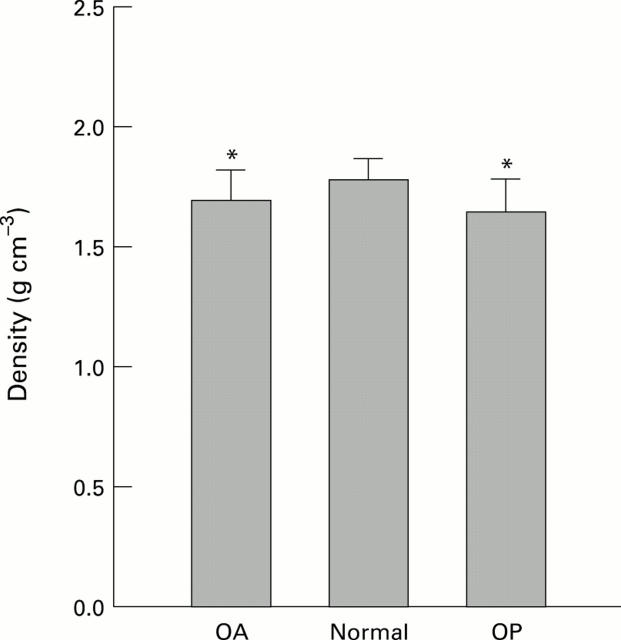
Results: Overall, OP bone was the least stiff and dense, followed by OA bone; normal bone was stiffer and more dense (p < 0.05). Though OP bone contained less mineral, the organic and water contents were increased in proportion suggesting no change in the relative amount of organic matrix. OA bone was also hypomineralised (p < 0.05) but had different organic and water fractions suggesting a defect in the matrix. Site variation of most properties was small, though across all the groups the superior region was significantly stiffer than the inferior.

Conclusion: This study shows that subchondral bone plate is less stiff than normal in both OP and OA and so cannot, by itself, explain the preserving of the overlying cartilage in OP while aiding its destruction in OA. However, the subchondral bone plate is only one part of the bony structure of the femoral head and changes in the cancellous bone need to be considered. The generalised changes in bone composition found in patients with OA support the hypothesis that the disease could involve the bone in the primary pathogenesis.


Figures

Figure 1
Figure 1
Schematic diagram of the sites on the human femoral head from which samples of subchondral bone were removed.
Figure 2
Figure 2
Median values of the stiffness of the subchondral bone plate determined from all sites over the femoral head of patients undergoing a hip replacement for osteoarthritis (OA), or osteoporosis (OP) compared with normal controls. The extent of the shaded bars shows the 25% and 75% confidence limits, the error bars show the 5% and 95% limits. (* p<0.05).
Figure 3
Figure 3
Density of the subchondral bone plate from the femoral heads of patients with OA or OP compared with normal controls. Both OA and OP groups are significantly reduced from normal and OP is slightly but significantly less than OA. (* p<0.05).
Figure 4
Figure 4
Composition of normal, OA, and OP subchondral bone plate from human femoral heads expressed as a fraction of the wet mass of the sample. Both OP and OA differed from normal but in different ways. (* p<0.05).
Figure 5
Figure 5
Composition of normal, OA, and OP subchondral bone plate expressed as mass per unit volume of tissue showing the changes seen in figure 4 in a different way. Both the OA and OP bone contain less mineral than normal (p < 0.05), though they have the same organic content. The increased water content in the OA bone matrix (p < 0.05 compared with normal) suggests that matrix changes occurring in the OA bone are different to those in OP bone.
Figure 6
Figure 6
Linear regression relations between stiffness and density of the subchondral bone plate. There is no difference between the gradients of the regression lines over the density range found in this study. See text for regression equations.
Figure 7
Figure 7
Site variation of stiffness of the subchondral bone plate from human femoral heads. (A) Stiffnesses are not normally distributed and the median values are shown. Shaded bars represent 25% and 75% and the errors bars the 5% and 95% confidence intervals. (B) Stiffness of each site expressed as a fractional change compared with the equivalent site from the normal group.
Figure 8
Figure 8
(A) Site variation of density of the subchondral bone plate from human femoral heads. (B) Density of each site expressed as a fractional change compared with the equivalent site from the normal group.

References

    1. Ann Rheum Dis. 1970 Jan;29(1):15-31 - PubMed
    1. Osteoarthritis Cartilage. 1995 Mar;3(1):25-33 - PubMed
    1. IEEE Trans Biomed Eng. 1970 Apr;17(2):101-5 - PubMed
    1. J Biomech. 1972 May;5(3):267-72 - PubMed
    1. Acta Orthop Scand. 1974;45(4):614-27 - PubMed

Publication types