. 1997 Apr;56(4):262-7.
doi: 10.1136/ard.56.4.262.

Characterisation of fibroblast-like cells in pannus lesions of patients with rheumatoid arthritis sharing properties of fibroblasts and chondrocytes

Affiliations

Characterisation of fibroblast-like cells in pannus lesions of patients with rheumatoid arthritis sharing properties of fibroblasts and chondrocytes

C Xue et al. Ann Rheum Dis. 1997 Apr.

Abstract

Objective: To better understand the characteristics of synoviocytes located in the rheumatoid arthritis (RA) pannus.

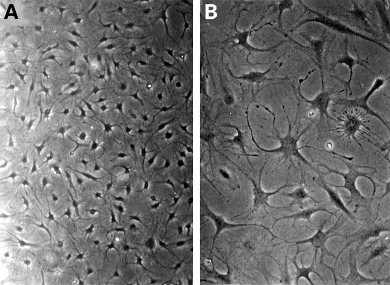
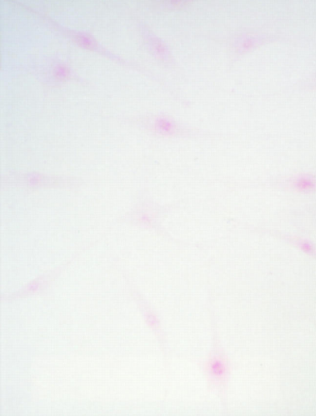
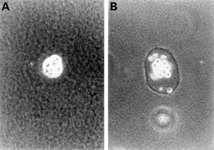
Methods: One cell line, termed PSC, was cloned from RA pannus lesions. Phenotypic analysis was done by contrast microscopy, indirect immunostaining, and safranin O staining. Transcription of several protooncogenes and matrix degrading enzymes was evaluated. The expression of mRNA for collagen II was detected by in situ hybridisation. The ability of anchorage independent growth was assessed by soft agarose culture.

Results: PSCs showed a high transcription of protooncogenes c-fos, c-myc and c-jun. They also expressed mRNA for matrix degrading enzymes, such as collagenase, cathepsin B, and cathepsin L. Anchorage independent growth assay demonstrated that PSCs formed colonies in soft agar culture. Phenotypic analysis showed that this fibroblast-like PSC was stained intensely with anti-vimentin and anti-fibroblast antibody. In situ reverse transcriptase assay showed that the cell line expressed type II collagen mRNA.

Conclusion: Alternative fibroblast-like cells were identified in the pannus lesion of RA sharing properties of fibroblasts and chondrocytes. These findings suggest that this fibroblast-like cell derived from pannus lesions may contribute to the destruction of the cartilage in RA.


Figures

Figure 1
Figure 1
Contrast microscopy of cultured pannus synoviocytes. PSCs were cultured in a 90 mm dish. They were a homogeneous group of cells (A) and had many filopodia (B) (original magnification × 200).
Figure 2
Figure 2
Immunofluorescence micrograph of vimentin filaments in cultured PSCs.Under contrast microscope, almost all PSCs were stained with anti-vimentin antibody labelled with FITC. The network of intermediate filament vimentin was stained with antibody to vimentin protein. An extensive network of vimentin filament surrounds the nucleus and extends out to the cell periphery (fluorescence microscopy, original magnification × 400).
Figure 3
Figure 3
Safranin O staining of synoviocytes. Cloned cells were seeded into the slide chamber, and fixed three days later with acetone, then subjected to the staining procedure described in the text. PSCs stained with red colour represent positive staining (original magnification × 200).
Figure 4
Figure 4
Type II collagen in situ reverse transcription. In situ reverse transcriptase assay was performed on the slide chamber with cultured PSCs to evaluate the expression of type II collagen mRNA. PCR amplification was performed with digoxigenin labelled oligonucleotide probe and sequential immunostaining with sheep anti-digoxigenin conjugated rhodamine. Cells picked up from the soft agar highly expressed type II collagen mRNA (A). High power view shows one PSC with several filopodia. Type II collagen mRNA shows a scattered pattern (B) (confocal microscopy, (A) original magnification × 200, (B) × 400).
Figure 5
Figure 5
Colony growth in anchorage independent growth assay. After culture for four weeks, PSCs formed colonies in soft agarose (A). Under the phase contrast microscope, a number of the colonies have a ring formed in the extracellular matrix (B) (contrast microscopy, (A) and (B) original magnification × 200).

References

    1. Cell. 1974 Dec;3(4):355-9 - PubMed
    1. Arthritis Rheum. 1995 Apr;38(4):477-84 - PubMed
    1. J Cell Sci. 1977 Aug;26:119-37 - PubMed
    1. Proc Natl Acad Sci U S A. 1977 Dec;74(12):5589-92 - PubMed
    1. Verh Dtsch Ges Pathol. 1980;64:193-212 - PubMed

Publication types

MeSH terms

Substances