. 1997 Jun 2;137(5):1001-16.
doi: 10.1083/jcb.137.5.1001.

Insertional mutation of the Drosophila nuclear lamin Dm0 gene results in defective nuclear envelopes, clustering of nuclear pore complexes, and accumulation of annulate lamellae

Affiliations

Insertional mutation of the Drosophila nuclear lamin Dm0 gene results in defective nuclear envelopes, clustering of nuclear pore complexes, and accumulation of annulate lamellae

B Lenz-Böhme et al. J Cell Biol. .

Abstract

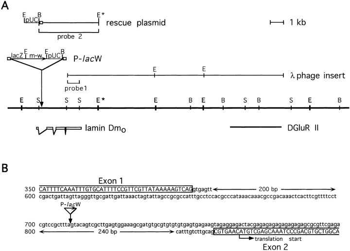
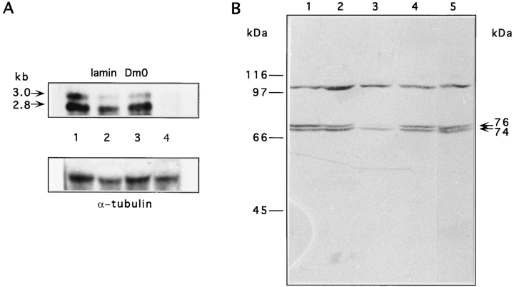
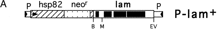
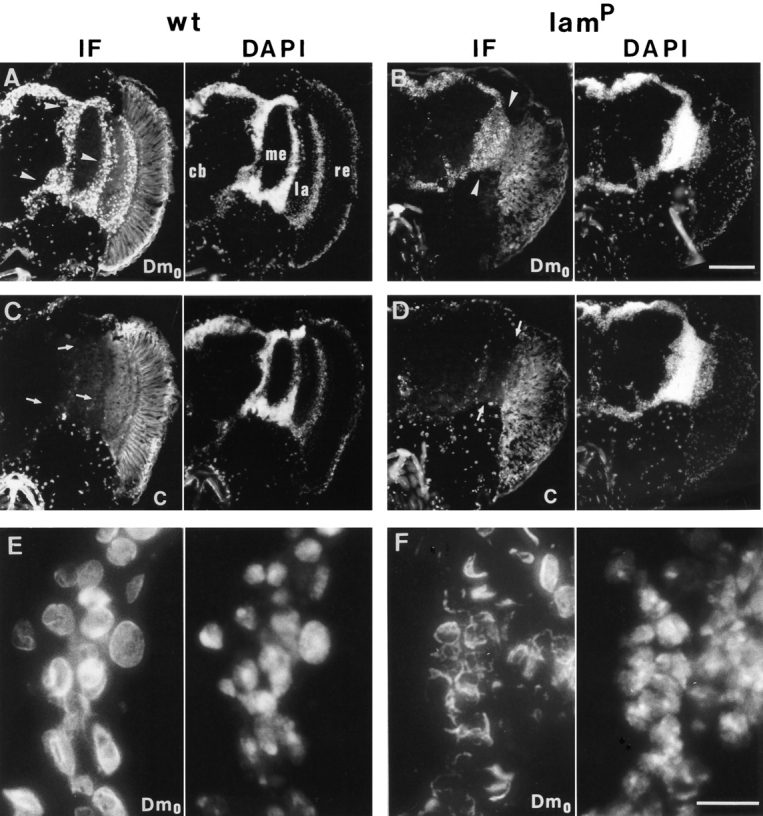
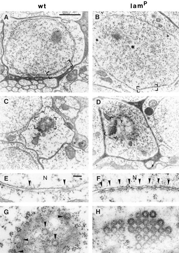
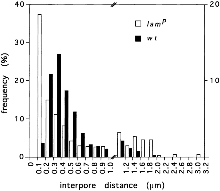
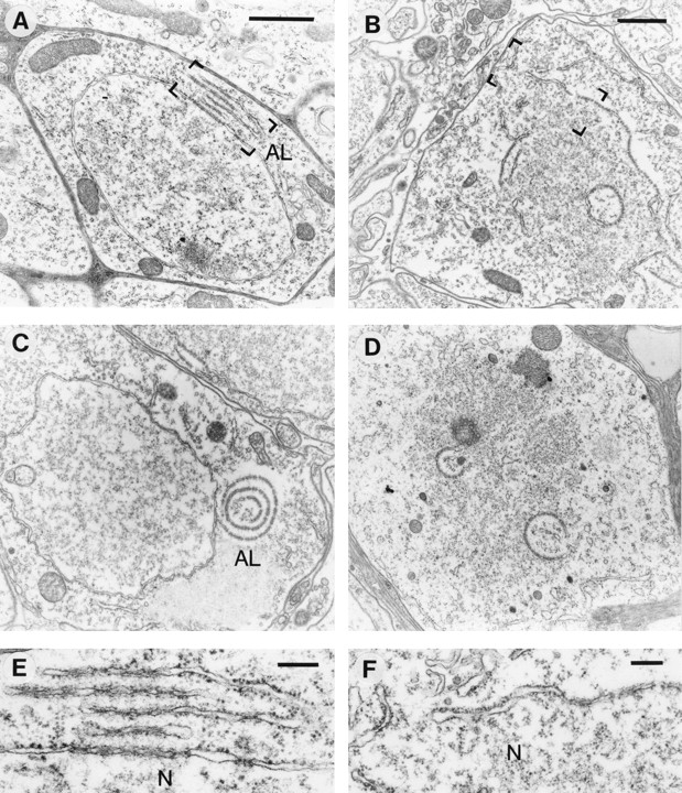
Nuclear lamins are thought to play an important role in disassembly and reassembly of the nucleus during mitosis. Here, we describe a Drosophila lamin Dm0 mutant resulting from a P element insertion into the first intron of the Dm0 gene. Homozygous mutant animals showed a severe phenotype including retardation in development, reduced viability, sterility, and impaired locomotion. Immunocytochemical and ultrastructural analysis revealed that reduced lamin Dm0 expression caused an enrichment of nuclear pore complexes in cytoplasmic annulate lamellae and in nuclear envelope clusters. In several cells, particularly the densely packed somata of the central nervous system, defective nuclear envelopes were observed in addition. All aspects of the mutant phenotype were rescued upon P element-mediated germline transformation with a lamin Dm0 transgene. These data constitute the first genetic proof that lamins are essential for the structural organization of the cell nucleus.


Figures

Figure 1
Figure 1
P-lacW insertion into the lamin Dm0 gene. (A) Schematic diagram of the genomic region at position 25F1-2 of the second chromosome covering the lamin Dm0 and the DGluR-II loci. Exonic regions of the lamin gene are indicated by open boxes below the restriction map. The restriction sites given are: E, EcoRI; B, BamHI; S, SalI. E* denotes a polymorphic EcoRI restriction site present in wt but not the w1118 strains. The P-lacW element is inserted into the first intron of the lamin gene (solid arrow). A horizontal arrow indicates the 5′ to 3′ orientation of the β-galactosidase (lacZ) and miniwhite (m-w) genes, and small boxes denote the inverted repeats of the P element. The λ phage insert, originally isolated for DGluR-II, extends into the lamin Dm0 gene region and hybridizes to the rescue plasmid derived from pUC sequences and adjacent genomic regions. Hybridization probes used for Northern (probe 1) and Southern (probe 2) blot analysis are indicated. (B) Precise location of the P-lacW insertion in the Dm0 genomic sequence. The P element is inserted into the center of the first intron of the lamin gene. Open boxes and capital letters indicate exonic regions. The translation start site is found in the second exon.
Figure 2
Figure 2
Expression of lamin Dm0 transcripts and proteins. (A) Northern blot of total RNA isolated from adult flies. The top panel shows hybridization with a lamin-specific riboprobe (Fig. 1 A, probe 1) and the lower with an α-tubulin riboprobe as control. Lane 1 contains total RNA of homozygous w1118, lane 2 of In(2LR)Gla, lane 3 of heterozygous lamP, and lane 4 of homozygous lamP flies. Arrows on the left indicate the sizes of the two lamin transcripts derived by alternative polyadenylation. Signals for both transcripts were very faint with the homozygous mutant strain (lane 4), but clearly visible after overexposure (not shown). (B) Western blot analysis of homogenates from single adult flies. The blot was probed with the monoclonal antibody U25 recognizing all Dm isoforms (Risau et al., 1981). Lane 1 contains the homogenate of a wt, lane 2 of a heterozygous lamP, lane 3 of a homozygous lamP, lane 4 of a homozygous Tw2-lamP, and lane 5 of a homozygous w1118 fly. Both lamin protein bands were reduced in the homozygous mutant (lane 3) and restored in the rescue fly (lane 4). A cross reacting protein band of apparent molecular mass of 105 kD is not related to the Dm0 gene product and served as an internal control for protein load. Positions of molecular weight marker proteins are indicated on the left; arrows on the right depict the positions of lamin isoforms.
Figure 3
Figure 3
Analysis of locomotor behavior and sterility. (A) Righting responses of wt, homo- and heterozygous lamP, and homozygous Tw2lamP flies. Six animals of each genotype were analyzed in six independent measurements. Mean values ± SEM of the observed righting times are given for each individual. (B and C) Histology of ovaries. Hematoxiline/eosin-stained paraffin sections of the ovaries from a wt (B) and a homozygous lamP (C) female fly are shown. Note shortened oocytes and reduced number of egg chambers in the bottom panel. ec, egg chamber; fc, follicle cell; g, germarium; gt, gut; nc, nurse cell; oc, oocyte. The scale bar in B represents 50 μm and is also valid for C.
Figure 3
Figure 3
Analysis of locomotor behavior and sterility. (A) Righting responses of wt, homo- and heterozygous lamP, and homozygous Tw2lamP flies. Six animals of each genotype were analyzed in six independent measurements. Mean values ± SEM of the observed righting times are given for each individual. (B and C) Histology of ovaries. Hematoxiline/eosin-stained paraffin sections of the ovaries from a wt (B) and a homozygous lamP (C) female fly are shown. Note shortened oocytes and reduced number of egg chambers in the bottom panel. ec, egg chamber; fc, follicle cell; g, germarium; gt, gut; nc, nurse cell; oc, oocyte. The scale bar in B represents 50 μm and is also valid for C.
Figure 4
Figure 4
Germline transformation with a lamin transgene. (A) Schematic diagram of the P element construct used for transformation. The Dm0 lamin transgene was inserted into the P transposable vector pHS85. Exonic regions of the lamin Dm0 gene (lam) are shown as black boxes. Restriction sites used for lamin transgene construction (see Materials and Methods) are indicated below: B, BamHI; M, MunI; EV, EcoRV. The P element transfer vector pHS85 contains a fusion of hsp82 protein (hatched) and neomycin phosphotransferase (stippled) gene. The hsp82 promoter is indicated by an arrow. Flanking P element sequences (P) are shown with their inverted repeats (triangles). (B) Southern blot analysis of the transformed rescue strain. EcoRI-restricted genomic DNA isolated from single flies was hybridized to a radioactively labeled plasmid rescue fragment (Fig. 1 A, probe 2). DNA samples were prepared from the following flies: hetero- and (lane 1), homozygous lamP (lane 2), heterozygous Tw2-lamP (one transgene copy, lane 3; two transgene copies, lane 4), homozygous Tw2-lamP (lane 5), and homozygous w1118 (lane 6). Arrows on the right indicate the control (9.8 kb), transgene (9.3 kb), and lamP (6.7 kb) specific hybridization bands.
Figure 4
Figure 4
Germline transformation with a lamin transgene. (A) Schematic diagram of the P element construct used for transformation. The Dm0 lamin transgene was inserted into the P transposable vector pHS85. Exonic regions of the lamin Dm0 gene (lam) are shown as black boxes. Restriction sites used for lamin transgene construction (see Materials and Methods) are indicated below: B, BamHI; M, MunI; EV, EcoRV. The P element transfer vector pHS85 contains a fusion of hsp82 protein (hatched) and neomycin phosphotransferase (stippled) gene. The hsp82 promoter is indicated by an arrow. Flanking P element sequences (P) are shown with their inverted repeats (triangles). (B) Southern blot analysis of the transformed rescue strain. EcoRI-restricted genomic DNA isolated from single flies was hybridized to a radioactively labeled plasmid rescue fragment (Fig. 1 A, probe 2). DNA samples were prepared from the following flies: hetero- and (lane 1), homozygous lamP (lane 2), heterozygous Tw2-lamP (one transgene copy, lane 3; two transgene copies, lane 4), homozygous Tw2-lamP (lane 5), and homozygous w1118 (lane 6). Arrows on the right indicate the control (9.8 kb), transgene (9.3 kb), and lamP (6.7 kb) specific hybridization bands.
Figure 5
Figure 5
Indirect immunofluorescence staining of head cryosections by lamin Dm0- and lamin C-specific monoclonal antibodies. Simultaneously processed head sections of a wt (left column) and a homozygous lamP (right column) fly are depicted. The left half column of each depicts the lamin antibody staining detected by indirect immunofluorescense (IF) and the right half the corresponding DNA staining by DAPI of the same section. A and B show lamin Dm0-specific staining by antibody ADL67 and C and D lamin C-specific staining by antibody LC28, of a total head hemisection each, with A and C as well as B and D, representing consecutive sections. E and F show magnifications of selected areas stained with antibody ADL67. Note the altered lamin Dm0 nuclear staining in lamP flies (B and F) as compared to wt (A and E). Arrowheads in A and B indicate the medulla and lobula/lobula plate cell body regions inspected in the electron-microscopic analysis. Arrows in C and D indicate the same areas displaying low lamin C expression. Anatomical structures of the fly's central nervous system are indicated in A: cb, central brain; me, medulla; la, lamina; re, retina. The retina displays strong autofluorescence, which is more obvious in wt. Bars: (A–D) 100 μm; (E and F) 10 μm.
Figure 6
Figure 6
Thin section, electron-microscopic analysis of cell bodies surrounding the medulla and lobula/lobula plate in wt, homo- and heterozygous lamP mutant, and in Tw2-lamP rescue flies. Electron micrographs of negatively stained sections are shown at lower magnification. A section through several optic lobe cell bodies of the homozygous (ho) lamP mutant (B) reveals striking differences to wt (A). Arrowheads in B point to examples of annulate lamellae (lower left cell) and NPC clusters in tangential (middle right cell) and transversal (lower right cell) nuclear sections. Similar sections from heterozygous (he) lamP (C) and homozygous Tw2-lamP (D) flies show no obvious differences to wt (A). Bar, 2 μm.
Figure 7
Figure 7
Distribution of NPCs in wt and homozygous lamP mutant cells. Electron micrographs representing thin sections from selected cells of the optic lobe region show NPCs in transversal (A and B) and tangential (C and D) nuclear sections. Bracketed areas (A–D) are shown at higher magnification in E–H, respectively. Arrowheads indicate NPCs in E–G. Note the high packing density of NPCs (NPC clusters) in B, D, F, and H with an apparent tetragonal symmetry in D and H. N, nucleoplasm. Bars: (A–D) 2 μm; (E–H) 100 nm.
Figure 8
Figure 8
Frequency of interpore distances of NPCs in wt and homozygous lamP mutant nuclei. Interpore distances between the NPCs of 30 cross sectioned nuclei, each of wt and lamP mutant cells determined from electron micrographs and classified into bin sizes of 0.1 μm (for distances ⩽1.0 μm) and 0.2 μm (for distances >1.0 μm), respectively. The graph shows the histogram of frequency (as a percentage) of wt (solid bars) and mutant (open bars) interpore distances (wt, n = 527; lamP, n = 529); scales differ on both sides of the figure, with the lamP values on the left and wt on the right. Note high frequency of short (<0.2 μm) interpore distances as well as an increased occurrence of long (>1.0 μm) interpore distances in the mutant nuclei.
Figure 9
Figure 9
Annulate lamellae and defective nuclear envelopes in homozygous lamP mutant cells. Thin-section electron micrographs show annulate lamellae (left column) and defective nuclear envelope (right column) structures typically found in mutant cells of the optic lobe region. A depicts a parallel stack of annulate lamellae (AL) close to an NPC cluster and (C) a circular annulate lamellae structure. B shows a cell with a partially fragmented nuclear membrane and D one without nuclear envelope. Bracketed areas of A and B are shown at higher magnification in E and F, respectively. N, Nucleoplasm. Bars: (A and B) 1 μm; (E and F) 200 nm; Bar in A is also valid for C and D.

References

    1. Aaronson RP, Blobel G. Isolation of pore complexes in association with a lamin. Proc Natl Acad Sci USA. 1975;72:1007–1011. - PMC - PubMed
    1. Aebi U, Cohn JB, Buhle L, Gerace L. The nuclear lamina is a meshwork of intermediate-type filaments. Nature (Lond) 1986;323:560–564. - PubMed
    1. Aitchison JD, Blobel G, Rout MP. Nup120p: a yeast nucleoporin required for NPC distribution and RNA transport. J Cell Biol. 1995;131:1659–1675. - PMC - PubMed
    1. Ashburner, M. 1989. Drosophila. A Laboratory Manual. Cold Spring Harbor Laboratory, Cold Spring Harbor, New York.
    1. Bailer SM, Eppenberger HM, Griffiths G, Nigg EA. Characterization of a 54-kD protein of the inner nuclear membrane: evidence for cell cycle–dependent interaction with the nuclear lamina. J Cell Biol. 1991;114:389–400. - PMC - PubMed

Publication types

MeSH terms