. 1997 Jun 2;137(5):1029-40.
doi: 10.1083/jcb.137.5.1029.

Intrinsic signals in the unique domain target p56(lck) to the plasma membrane independently of CD4

Affiliations

Intrinsic signals in the unique domain target p56(lck) to the plasma membrane independently of CD4

M J Bijlmakers et al. J Cell Biol. .

Abstract

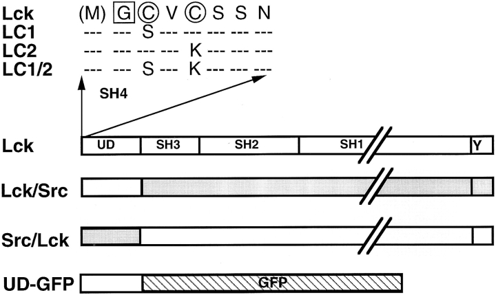
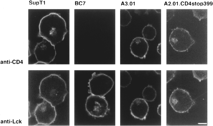
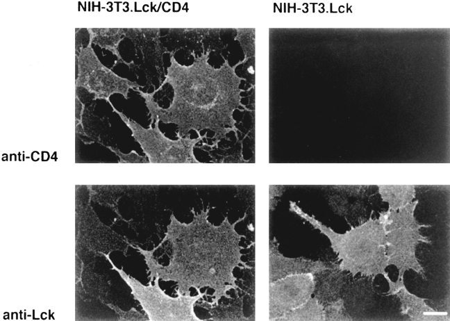
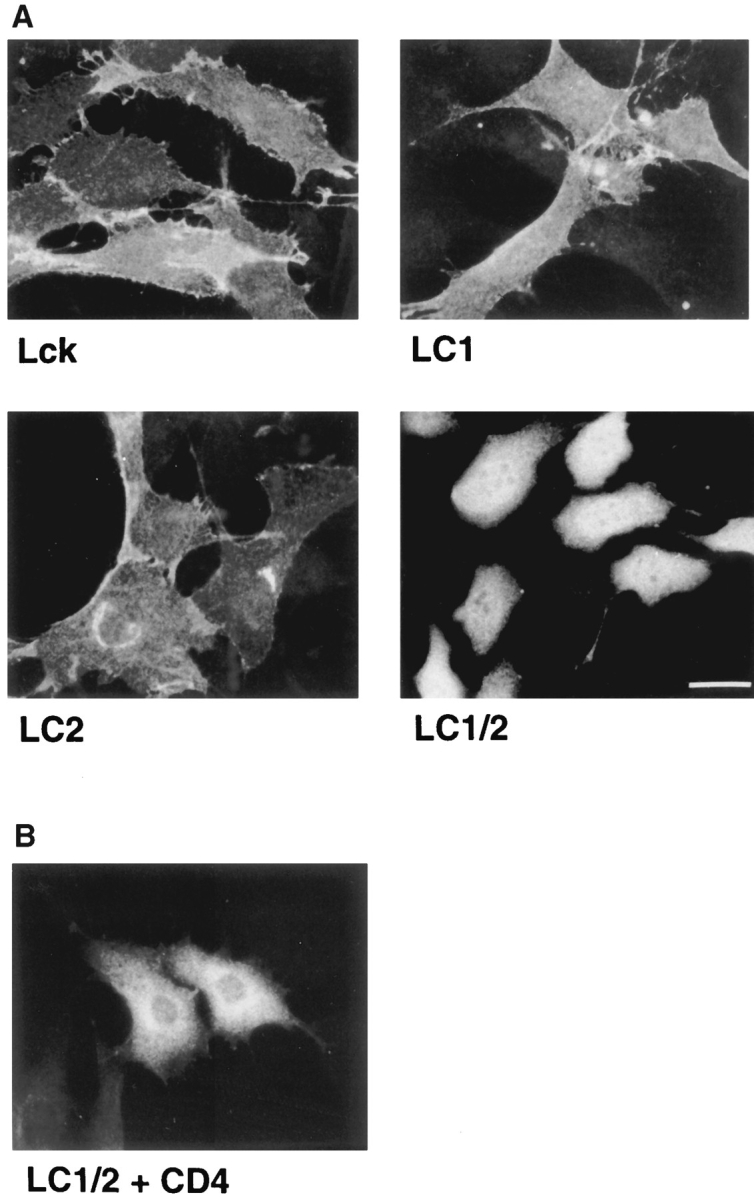
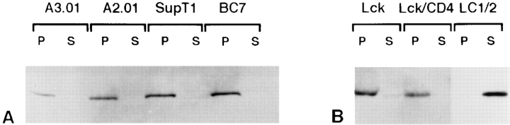
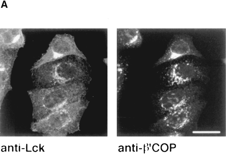
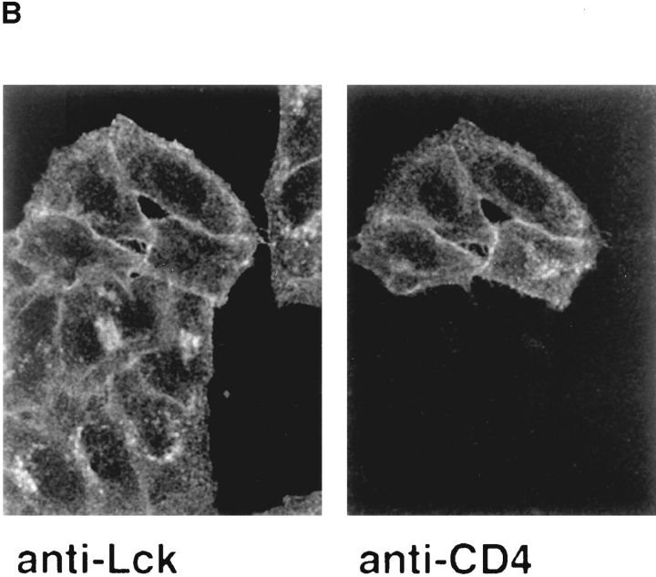
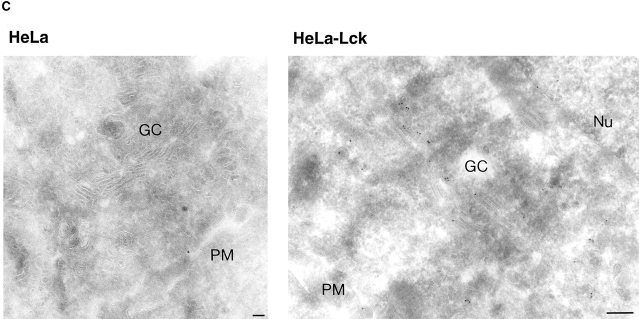
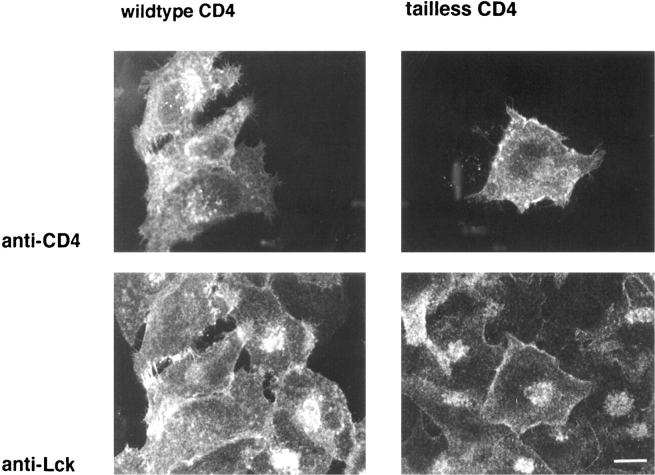
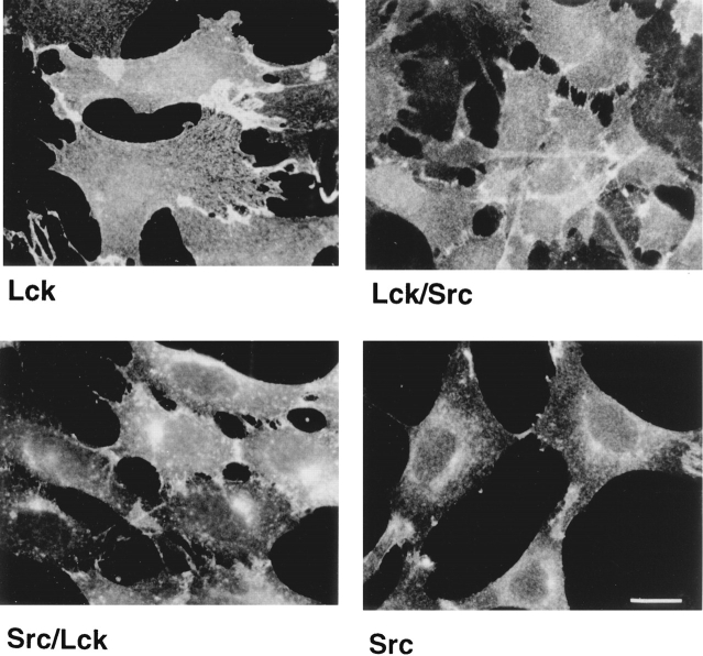
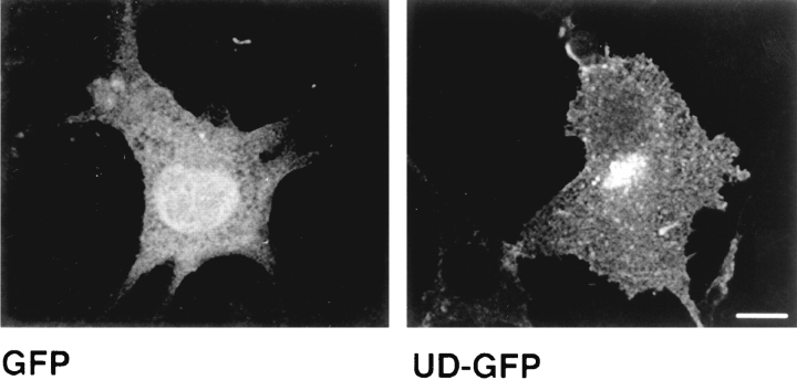
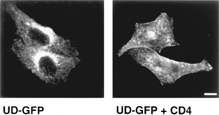
In T lymphocytes, the Src-family protein tyrosine kinase p56(lck) (Lck) is mostly associated with the cytoplasmic face of the plasma membrane. To determine how this distribution is achieved, we analyzed the location of Lck in lymphoid and in transfected nonlymphoid cells by immunofluorescence. We found that in T cells Lck was targeted correctly, independently of the cell surface proteins CD4 and CD8 with which it interacts. Similarly, in transfected NIH-3T3 fibroblasts, Lck was localized at the plasma membrane, indicating that T cell-specific proteins are not required for targeting. Some variation in subcellular distribution was observed when Lck was expressed in HeLa and MDCK cells. In these cells, Lck associated with both the plasma membrane and the Golgi apparatus, while subsequent expression of CD4 resulted in the loss of Golgi-associated staining. Together, these data indicate that Lck contains intrinsic signals for targeting to the plasma membrane. Furthermore, delivery to this site may be achieved via association with exocytic transport vesicles. A mutant Lck molecule in which the palmitoylation site at cysteine 5 was changed to lysine (LC2) localized to the plasma membrane and the Golgi region in NIH3T3 cells. However, the localization of a mutant in which the palmitoylation site at cysteine 3 was changed to serine (LC1) was indistinguishable from wild-type Lck. Chimeras composed of only the unique domain of Lck linked to either c-Src or the green fluorescent protein similarly localized to the plasma membrane of NIH-3T3 cells. Thus, the targeting of Lck appears to be determined primarily by its unique domain and may be influenced by the use of different palmitoylation sites.


Figures

Figure 1
Figure 1
Domain organization of the Src-family proteins and of the Lck constructs used in this study. Lck is shown schematically as a representative of Src-family proteins. Indicated are the unique domain (UD), the Src Homology 3 (SH3), SH2, the tyrosine kinase (SH1) domains, and the short conserved COOHterminal region that contains the “regulatory” tyrosine (Y). The amino acid sequence of the conserved NH2-terminal region, also designated the SH4 domain, which contains the signals for acylation is shown for Lck. The attachment of myristic acid to glycine (boxed) requires the removal of the NH2-terminal methionine and the consensus sequence GXXXS/C. Palmitic acid is attached to cysteines (encircled) and requires a myristoylated NH2-terminal glycine and cysteine on position 3. Cysteine 5 in Lck is also a palmitate acceptor site.In this study we used Lck mutants with disruptions to the first (LC1), the second (LC2), or both (LC1/2) palmitoylation sites (Turner et al., 1990). Also depicted are the chimeras used in this study: Lck/Src contains the unique domain of Lck and the remainder of Src, Src/Lck contains the unique domain of Src and the remainder of Lck (Turner et al., 1990). The chimera UD-GFP is composed of the unique domain of Lck fused to the NH2 terminus of green fluorescent protein.
Figure 2
Figure 2
Distribution of Lck in T cells. Double indirect immunofluorescence staining of Lck and CD4 in the CD4-expressing T cell line SupT1 and its CD4-negative derivative, BC7, as well as for the CD4-positive T cell line A3.01 and its derivative A2.01-CD4stop399, that expresses CD4 without a cytoplasmic tail. Rabbit anti-Lck (Lck I) was detected with FITC-conjugated goat anti–rabbit antibodies, murine anti-CD4 (Q4120) with rhodamine-conjugated goat anti–mouse antibodies. Observation was by confocal microscopy of 3-μm-thick optical sections. Bar, 12.5 μm.
Figure 3
Figure 3
Distribution of Lck in NIH-3T3 fibroblasts. Double indirect immunofluorescence staining of Lck and CD4 in fibroblasts either stably transfected with both CD4 and murine Lck (left) or with murine Lck alone (right). Primary and secondary antibodies were as described in the legend to Fig. 1. Optical sections (3 μm) were observed by confocal microscopy. Bar, 20 μm.
Figure 10
Figure 10
Cellular distribution of Lck palmitoylation mutants in NIH-3T3 cells. (A) NIH-3T3 cells were stably transfected with either wild-type Lck, LC1, LC2, or LC1/2 (see Fig. 1). Cells were stained by indirect immunofluorescence with an antibody against Lck (Lck I). (B) CD4-expressing NIH-3T3 cells were transfected with LC1/2 and stained for Lck with Lck I. Cells were observed by fluorescence microscopy. Bar, 25 μm.
Figure 4
Figure 4
Membrane association of Lck in the presence and absence of CD4. Cells were broken by passage through a ball-bearing homogenizer. After removal of unbroken cells and nuclei, cellular membranes were recovered by centrifugation at 100,000 g (P, pellet) and separated from the soluble fraction (S, supernatant). For each cell line, samples representing equivalent amounts of cells were analyzed by SDS-PAGE and subsequent immunoblotting with the affinity-purified anti-Lck antibodies (Lck II). (A) T cell lines expressing CD4 (SupT1 and A3.01), tailless CD4 (A2.01) or no CD4 (BC7). (B) NIH-3T3 cells transfected with Lck alone, with both Lck and CD4, or with the palmitoylation mutant LC1/2.
Figure 5
Figure 5
Distribution of Lck in HeLa cells. (A) HeLa cells stably expressing murine Lck were double-stained with antibodies against Lck (Lck I) and β′-COP. Lck staining was detected with FITC-conjugated goat anti–rabbit antibodies, β′-COP staining with rhodamineconjugated goat anti–rat antibodies. (B) HeLa cells stably expressing human Lck were transfected with a cDNA encoding CD4. 2 d later, the cells were double-stained for Lck and CD4 (as in Fig. 1). Note the difference in distribution of Lck between cells that express CD4 (the four cells at the top) and cells that do not. Confocal images of a 3-μm optical section are shown. Bar, 20 μm. (C) Frozen thin sections of untransfected HeLa cells (left) or HeLa cells transfected with human Lck (right) were labeled with an antibody against Lck (Lck I) and as a second layer goat anti–rabbit IgG–conjugated gold (10 nm). Nu, nucleus; GC, Golgi complex; PM, plasma membrane. Bar, 0.1 μm.
Figure 5
Figure 5
Distribution of Lck in HeLa cells. (A) HeLa cells stably expressing murine Lck were double-stained with antibodies against Lck (Lck I) and β′-COP. Lck staining was detected with FITC-conjugated goat anti–rabbit antibodies, β′-COP staining with rhodamineconjugated goat anti–rat antibodies. (B) HeLa cells stably expressing human Lck were transfected with a cDNA encoding CD4. 2 d later, the cells were double-stained for Lck and CD4 (as in Fig. 1). Note the difference in distribution of Lck between cells that express CD4 (the four cells at the top) and cells that do not. Confocal images of a 3-μm optical section are shown. Bar, 20 μm. (C) Frozen thin sections of untransfected HeLa cells (left) or HeLa cells transfected with human Lck (right) were labeled with an antibody against Lck (Lck I) and as a second layer goat anti–rabbit IgG–conjugated gold (10 nm). Nu, nucleus; GC, Golgi complex; PM, plasma membrane. Bar, 0.1 μm.
Figure 6
Figure 6
Microinjection of wild-type CD4 and tailless CD4 into Hela-Lck cells. HeLa cells stably expressing human Lck were microinjected with cDNA for wildtype CD4 (left) or tailless CD4 (right). Cells were fixed 3 h after injection and stained with antibodies against Lck and CD4 (as in Fig. 1). Note the difference in distribution of Lck between cells that were injected with wild-type CD4 cDNA (left, the three cells on the left) and cells that were not injected. Also note that injection with tailless CD4 cDNA has no effect on the distribution of Lck (right, the cell in the middle). Confocal images of 3-μm optical sections are shown. Bar, 15 μm.
Figure 7
Figure 7
Cellular distribution of chimeras between Lck and Src. NIH-3T3 fibroblasts were stably transfected either with Lck, c-Src, Lck/Src, or Src/Lck as indicated. Src/Lck contains the unique domain of c-Src and the remainder of Lck, while Lck/Src contains the unique domain of Lck and the remainder of c-Src (see Fig. 1). Lck- and Src/Lck-expressing cells were stained with Lck I, and Src- and Lck/Src-expressing cells were stained with the anti-Src mAb 327. Cells were observed by fluorescence microscope. Bar, 20 μm.
Figure 8
Figure 8
Cellular distribution of GFP and UDGFP in NIH-3T3 cells. NIH-3T3 cells were transfected either with GFP or UD-GFP, a chimera composed of the unique domain of Lck linked to GFP. Cells were fixed 2 d later and observed by fluorescent confocal microscopy. Note the plasma membrane distribution of UD-GFP as opposed to the cytosolic distribution of GFP. Confocal images of 3-μm optical sections are shown. Bar, 15 μm.
Figure 9
Figure 9
Cellular distribution of UD-GFP in HeLa cells. UD-GFP was transfected into HeLa cells (left) or into HeLa cells that express CD4 (right). Cells were fixed 2 d later and 3-μm optical sections were observed by confocal microscopy. Bar, 10 μm.

References

    1. Alland L, Peseckis SM, Atherton RE, Berthiaume L, Resh MD. Dual myristylation and palmitylation of Src family member p59fyn affects subcellular localization. J Biol Chem. 1994;24:16701–16705. - PubMed
    1. August A, Dupont B. Activation of Src family kinase Lck following CD28 crosslinking in the Jurkat leukemic cell line. Biochem Biophys Res Commun. 1994;199:1466–1473. - PubMed
    1. Bar SD, Rotin D, Batzer A, Mandiyan V, Schlessinger J. SH3 domains direct cellular localization of signaling molecules. Cell. 1993;74:83–91. - PubMed
    1. Bell GM, Bolen JB, Imboden JB. Association of Src-like protein tyrosine kinases with the CD2 cell surface molecule in rat T lymphocytes and natural killer cells. Mol Cell Biol. 1992;12:5548–5554. - PMC - PubMed
    1. Berthiaume L, Resh MD. Biochemical characterization of a palmitoyl acyltransferase activity that palmitoylates myristoylated proteins. J Biol Chem. 1995;270:22399–22405. - PubMed

Publication types

MeSH terms