. 1997 Jun 2;137(5):1057-68.
doi: 10.1083/jcb.137.5.1057.

A natural hepatocyte growth factor/scatter factor autocrine loop in myoblast cells and the effect of the constitutive Met kinase activation on myogenic differentiation

Affiliations

A natural hepatocyte growth factor/scatter factor autocrine loop in myoblast cells and the effect of the constitutive Met kinase activation on myogenic differentiation

S Anastasi et al. J Cell Biol. .

Abstract

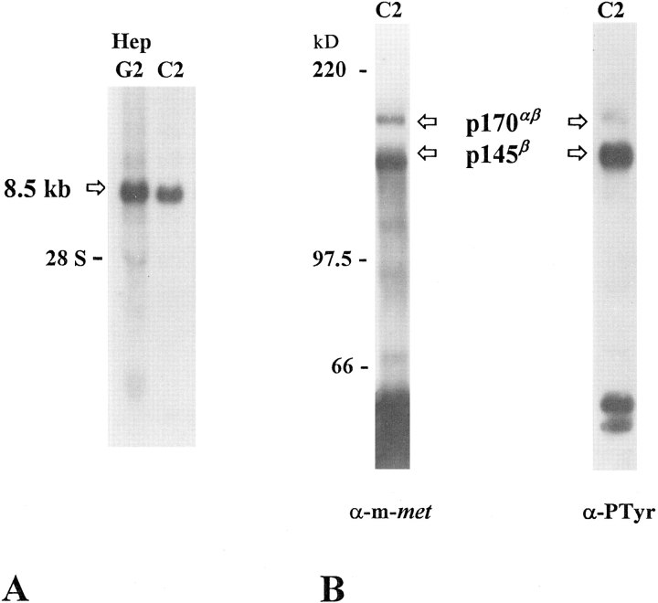
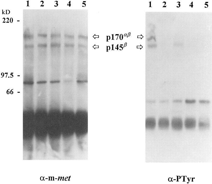
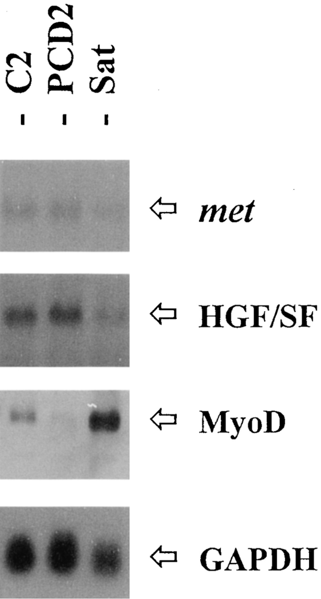
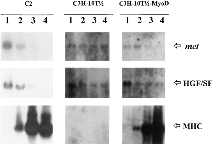
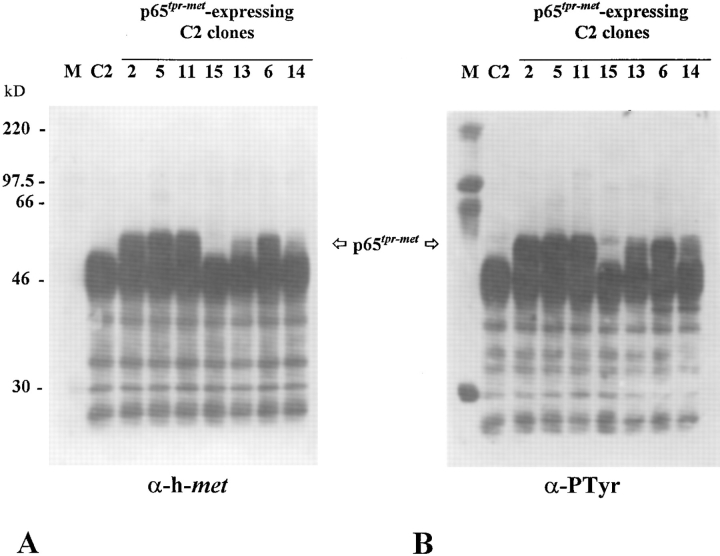
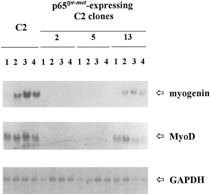
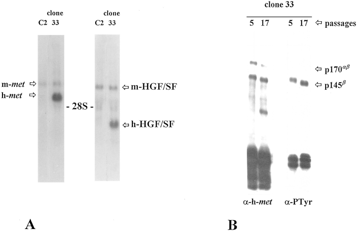
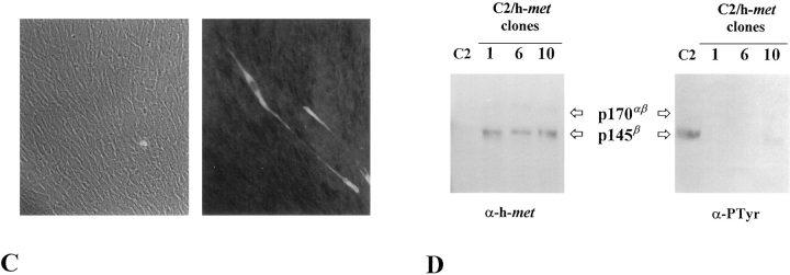
As a rule, hepatocyte growth factor/scatter factor (HGF/SF) is produced by mesenchymal cells, while its receptor, the tyrosine kinase encoded by the met proto-oncogene, is expressed by the neighboring epithelial cells in a canonical paracrine fashion. In the present work we show that both HGF/SF and met are coexpressed by undifferentiated C2 mouse myoblasts. In growing cells, the autocrine loop is active as the receptor exhibits a constitutive phosphorylation on tyrosine that can be abrogated by exogenously added anti-HGF/SF neutralizing antibodies. The transcription of HGF/SF and met genes is downregulated when myoblasts stop proliferating and differentiate. The coexpression of HGF/SF and met genes is not exclusive to C2 cells since it has been assessed also in other myogenic cell lines and in mouse primary satellite cells, suggesting that HGF/SF could play a role in muscle development through an autocrine way. To analyze the biological effects of HGF/SF receptor activation, we stably expressed the constitutively activated receptor catalytic domain (p65(tpr-met)) in C2 cells. This active kinase determined profound changes in cell shape and inhibited myogenesis at both morphological and biochemical levels. Notably, a complete absence of muscle regulatory markers such as MyoD and myogenin was observed in p65(tpr-met) highly expressing C2 clones. We also studied the effects of the ectopic expression of human isoforms of met receptor (h-met) and of HGF/SF (h-HGF/SF) in stable transfected C2 cells. Single constitutive expression of h-met or h-HGF/SF does not alter substantially the growth and differentiation properties of the myoblast cells, probably because of a species-specific ligand-receptor interaction. A C2 clone expressing simultaneously both h-met and h-HGF/SF is able to grow in soft agar and shows a decrease in myogenic potential comparable to that promoted by p65(tpr-met) kinase. These data indicate that a met kinase signal released from differentiation-dependent control provides a negative stimulus for the onset of myogenic differentiation.


Figures

Figure 1
Figure 1
Synthesis of met receptor by C2 myoblast cells. (A) Northern blot of total RNA extracted from human hepatocarcinoma HepG2 cells and C2 cells probed with human full-length met cDNA (Ponzetto et al., 1991). The position of 8.5-kb metrelated transcript and the 28S ribosomal RNA is indicated. (B) Activation state of met receptor expressed by C2 myoblasts. Protein extracts from proliferating C2 myoblasts were immunoprecipitated with anti–met rabbit SP260 antiserum (directed against the COOH-terminal portion of the murine protein), electrophoresed onto 8% reducing SDS-PAGE, and immunoblotted with SP260 antiserum (α-m-met). In these conditions, both the 145-kD β subunit of mature met receptor (p145β) and the 170-kD single-chain precursor (p170αβ) were detected. The same filter was stripped and reprobed with monoclonal anti-phosphotyrosine antibodies (α-PTyr) to provide evidence for the activation degree of p145β.
Figure 2
Figure 2
Synthesis of HGF/ SF by C2 myoblast cells. (A) Northern blot analysis of total RNA extracted from human hepatocarcinoma cells (HepG2) and C2 cells to illustrate the production of a 6-kb HGF/SF mRNA. As previously reported in literature (Shiota et al., 1992), HepG2 cells are negative for the expression of HGF/SF gene. (B and C) Phase-contrast micrographs of a scatter assay carried out with MDCK epithelial cells incubated with C2 myoblast–conditioned medium (B) or standard medium (C).
Figure 3
Figure 3
HGF/SF is an autocrine factor for C2 myoblasts. C2 proliferating cells exposed to culture conditions that remove HGF/SF molecules from cell surface (see Materials and Methods) were lysed, immunoprecipitated with anti–murine met antibodies, and analyzed for the tyrosine phosphorylation degree of HGF/SF receptor by Western blotting with anti-phosphotyrosine antibodies (α-PTyr). Control cells (lane 1); C2 cells acid treated and incubated with 0.5 M NaCl-containing DME (lane 2); cells treated as in lane 2 and further incubated with C2-conditioned medium (lane 3) or C2-conditioned medium pretreated with an antiHGF/SF neutralizing antibody (lane 4); cells as in lane 2 and further incubated with fresh 10% FCS-containing medium (lane 5).
Figure 4
Figure 4
Coexpression of met and HGF/SF in other myogenic cells. Total RNA from C2 myoblasts, PCD2 mouse myoblasts, and mouse primary satellite (Sat) cell cultures was hybridized with the indicated probes.
Figure 5
Figure 5
The expression of met and HGF/SF genes is downregulated concomitantly with the onset of myogenic differentiation. C2, C3H-10T1/2, and C3H-10T1/2-MyoD cells were cultured in DME containing 20% FCS up to confluency, and then transferred to low serum–containing medium (2% FCS). Total RNA extracted before serum deprivation (lanes 1) and 1 (lanes 2), 2 (lanes 3), and 3 (lanes 4) d after serum deprivation was assayed for the expression of met, HGF/SF, and a muscle-specific marker, MHC, transcripts. Quantitative decrease of met and HGF/SF mRNA synthesis was observed only in the presence of muscle differentiation.
Figure 6
Figure 6
Extensive p65tpr-met kinase activation in p65tpr-met- expressing C2 clones. Total cell lysates were immunoprecipitated with rabbit anti–met C12 antiserum (directed against the COOHterminal portion of the human protein), electrophoresed onto 12% reducing SDS-PAGE, and immunoblotted with the same antibody (A) or anti-phosphotyrosine antibodies (B). A good correlation was found between the p65tpr-met protein levels and its degree of tyrosine phosphorylation. Clone 15 was negative for the expression of p65tpr-met. Size in kD was estimated using a molecular mass standard (M).
Figure 7
Figure 7
Phenotypic appearance and differentiating properties of p65tpr-met-expressing C2 clones. Micrographs of p65tpr-met highly expressing cells (B) and C2 parental cells (A) cultured either in growth or differentiation medium. The myogenic potential was assessed by detecting myotube formation and by immunofluorescence staining for MHC in 48-h differentiation medium–exposed cell cultures.
Figure 8
Figure 8
Decreased expression of muscle-regulatory genes in p65tpr-met-expressing C2 clones. Total RNA from proliferating (lanes 1) and differentiating (lanes 2, 3, and 4; 24-, 48-, and 72-h postserum deprivation, respectively) cultures was hybridized with the indicated probes. To estimate if equal amounts of RNA were loaded, the same filter was probed with a constitutively expressed gene (GAPDH).
Figure 9
Figure 9
Double ectopic expression of h-met and h-HGF/ SF is required for the constitutive activation of h-met kinase. (A) Northern blot analysis of RNA expression levels of h-met and h-HGF/SF in clone 33 selected from C2 h-met/h-HGF/SF double transfection. Exogenous transcripts are larger than endogenous ones. (B) The h-met receptor expressed by clone 33 is phosphorylated on tyrosine residues. Cells were lysed at two different culture passages and immunoprecipitated with anti–human met antibodies. Proteins were resolved by 8% reducing SDSPAGE and immunoblotted with anti–human met (α-hmet) or anti-phosphotyrosine (α-PTyr) antibodies. (C) Phase-contrast and anti-MHC immunofluorescence micrographs of clone 33 cells exposed for 48 h in differentiation medium containing 10% HS. (D) Cell lysates from C2 parental cells and three single h-met–expressing C2 (C2/ h-met) clones were immunoprecipitated with anti–murine met antiserum (C2 cells) or with anti-human met antiserum (C2/h-met clones), respectively. The immunocomplexes were resolved by 8% reducing SDS-PAGE and immunoblotted with anti–human (α-h-met) or anti-phosphotyrosine mAbs (α-PTyr). The almost undetectable tyrosine phosphorylation exhibited by h-met in comparison with the endogenous receptor suggests that it is not efficiently activated by murine HGF/SF.
Figure 9
Figure 9
Double ectopic expression of h-met and h-HGF/ SF is required for the constitutive activation of h-met kinase. (A) Northern blot analysis of RNA expression levels of h-met and h-HGF/SF in clone 33 selected from C2 h-met/h-HGF/SF double transfection. Exogenous transcripts are larger than endogenous ones. (B) The h-met receptor expressed by clone 33 is phosphorylated on tyrosine residues. Cells were lysed at two different culture passages and immunoprecipitated with anti–human met antibodies. Proteins were resolved by 8% reducing SDSPAGE and immunoblotted with anti–human met (α-hmet) or anti-phosphotyrosine (α-PTyr) antibodies. (C) Phase-contrast and anti-MHC immunofluorescence micrographs of clone 33 cells exposed for 48 h in differentiation medium containing 10% HS. (D) Cell lysates from C2 parental cells and three single h-met–expressing C2 (C2/ h-met) clones were immunoprecipitated with anti–murine met antiserum (C2 cells) or with anti-human met antiserum (C2/h-met clones), respectively. The immunocomplexes were resolved by 8% reducing SDS-PAGE and immunoblotted with anti–human (α-h-met) or anti-phosphotyrosine mAbs (α-PTyr). The almost undetectable tyrosine phosphorylation exhibited by h-met in comparison with the endogenous receptor suggests that it is not efficiently activated by murine HGF/SF.

References

    1. Adams JC, Furlong RA, Watt FM. Production of scatter factor by ndk, a strain of epithelial cells, and inhibition of scatter factor activity by suramin. J Cell Sci. 1991;98:385–394. - PubMed
    1. Allen RE, Sheehan SM, Taylor RG, Kendall TL, Rice GM. Hepatocyte growth factor activates quiescent skeletal muscle satellite cells in vitro. J Cell Physiol. 1995;165:307–312. - PubMed
    1. Bader D, Masaki T, Fischman DA. Immunochemical analysis of myosin heavy chain during avian myogenesis in vivo and in vitro. J Cell Biol. 1982;95:763–770. - PMC - PubMed
    1. Bellusci S, Moens G, Gaudino G, Comoglio PM, Nakamura T, Thiery J-P, Jouanneau J. Creation of an hepatocyte growth factor/scatter factor autocrine loop in carcinoma cells induces invasive properties associated with increased tumorigenicity. Oncogene. 1994;9:1091–1099. - PubMed
    1. Bladt F, Riethmacher D, Isenmann S, Aguzzi A, Birchmeier C. Essential role for the c-metreceptor in the migration of myogenic precursor cells into the limb bud. Nature (Lond) 1995;376:768–771. - PubMed

Publication types

MeSH terms