. 1997 Jun 2;185(11):1909-18.
doi: 10.1084/jem.185.11.1909.

Human dendritic cells skew isotype switching of CD40-activated naive B cells towards IgA1 and IgA2

Affiliations

Human dendritic cells skew isotype switching of CD40-activated naive B cells towards IgA1 and IgA2

J Fayette et al. J Exp Med. .

Abstract

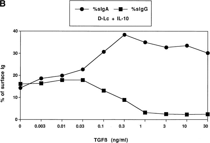
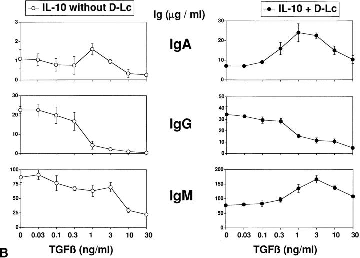
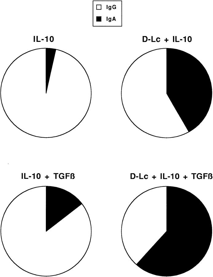
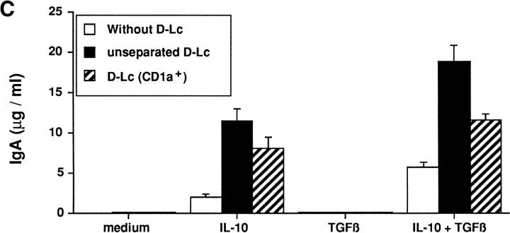
Within T cell-rich areas of secondary lymphoid organs, interdigitating dendritic cells recruit antigen-specific T cells that then induce B cells to secrete Igs. This study investigates the possible role(s) of dendritic cells in the regulation of human B cell responses. In the absence of exogenous cytokines, in vitro generated dendritic cells (referred to as Dendritic Langerhans cells, D-Lc) induced surface IgA expression on approximately 10% of CD40-activated naive sIgD+ B cells. In the presence of IL-10 and TGF-beta, a combination of cytokines previously identified for its capacity to induce IgA switch, D-Lc strongly potentiated the induction of sIgA on CD40-activated naive B cells from 5% to 40-50%. D-Lc alone did not induce the secretion of IgA by CD40-activated naive B cells, which required further addition of IL-10. Furthermore, D-Lc skewed towards the IgA isotype at the expense of IgG, the Ig production of CD40-activated naive B cells cultured in the presence of IL-10 and TGF-beta. Importantly, under these culture conditions, both IgA1 and IgA2 were detected. In the presence of IL-10, secretion of IgA2 by CD40-activated naive B cells could be detected only in response to D-Lc and was further enhanced by TGF-beta. Collectively, these results suggest that in addition to activating T cells in the extrafollicular areas of secondary lymphoid organs, human D-Lc also directly modulate T cell-dependent B cell growth and differentiation, by inducing the IgA isotype switch.


Figures

Figure 1
Figure 1
Surface IgA1 and IgA2 labelings are specific. Cells (5 × 10⁵) from a sIgA1+ EBV clone (VLMB 325) and from a sIgA2+ EBV clone (HEL) were stained, respectively, with FITC-labeled anti-sIgA1 and -sIgA2 mAbs (dotted lines). Competition experiments were done by preincubating FITC-labeled anti-sIgA1 or anti-sIgA2 (right) together with 50 μg/ml human IgA1λ + κ myelomas (left) or with 50 μg/ml human IgA1λ + κ myelomas, which were then used to stain each EBV clone (black lines). Controls with unrelated mAb of the same isotype were overlapped in each panel (gray histograms).
Figure 2
Figure 2
In vitro generated D-Lc enhance CD40-activated naive B cell proliferation. 5 × 10⁴/ml highly purified sIgD+ B lymphocytes were cultured in a final volume of 200 μl over 1.25 × 10⁴/ml irradiated CD40L-L cells in the absence or with increasing numbers of D-Lc (irradiated or not) without exogenous cytokine. Thymidine uptake, determined after 6 d (A), or at different indicated time points (B), is expressed as mean ± SD of triplicate cultures. (C) DNA synthesis of 5 × 10⁴/ml CD40-activated sIgD+ B cultured 6 d in the absence or presence of 5 × 10⁴/ml in vitro generated D-Lc with or without IL-10 (200 ng/ml), TGF-β (0.3 ng/ml), or both. (D) For enumeration experiments, 5 × 10⁴/ml sIgD+ B were cultured for 6 d in the presence or absence of 5 × 10⁴/ml D-Lc, with or without IL-10 (200 ng/ml) and increasing doses of TGF-β (0.1, 0.3, 1, and 3 ng/ml). Viable cells that were not colored with Trypan blue dye were enumerated at day 6. One representative experiment is shown out of 4 in A and B, 6 in C, and 2 in D.
Figure 2
Figure 2
In vitro generated D-Lc enhance CD40-activated naive B cell proliferation. 5 × 10⁴/ml highly purified sIgD+ B lymphocytes were cultured in a final volume of 200 μl over 1.25 × 10⁴/ml irradiated CD40L-L cells in the absence or with increasing numbers of D-Lc (irradiated or not) without exogenous cytokine. Thymidine uptake, determined after 6 d (A), or at different indicated time points (B), is expressed as mean ± SD of triplicate cultures. (C) DNA synthesis of 5 × 10⁴/ml CD40-activated sIgD+ B cultured 6 d in the absence or presence of 5 × 10⁴/ml in vitro generated D-Lc with or without IL-10 (200 ng/ml), TGF-β (0.3 ng/ml), or both. (D) For enumeration experiments, 5 × 10⁴/ml sIgD+ B were cultured for 6 d in the presence or absence of 5 × 10⁴/ml D-Lc, with or without IL-10 (200 ng/ml) and increasing doses of TGF-β (0.1, 0.3, 1, and 3 ng/ml). Viable cells that were not colored with Trypan blue dye were enumerated at day 6. One representative experiment is shown out of 4 in A and B, 6 in C, and 2 in D.
Figure 3
Figure 3
D-Lc induce CD40-activated naive B cells to express sIgA. 5 × 10⁴/ml sIgD+ B lymphocytes were cultured in a final volume of 1 ml in the absence or presence of 5 × 10⁴/ml D-Lc over 1.25 × 10⁴/ml irradiated CD40L-L cells without exogenous cytokine or with IL-10 (200 ng/ ml), TGF-β (0.3 ng/ml) or both (A), or with IL-10 and increasing doses of TGF-β (B). B cells were harvested after 7 d of culture and stained with rabbit polyclonal FITC-labeled anti-IgA or anti-IgG antibodies (A, B) or FITC-labeled anti-IgA1 or -IgA2 mAbs (C) and analyzed by FACScan®. Dead cells which incorporated propidium iodide were excluded. Histograms are presented (A) or percentages of sIgG and sIgA positively stained cells (B). One representative experiment out of six (A) and one out of three (B) are shown.
Figure 3
Figure 3
D-Lc induce CD40-activated naive B cells to express sIgA. 5 × 10⁴/ml sIgD+ B lymphocytes were cultured in a final volume of 1 ml in the absence or presence of 5 × 10⁴/ml D-Lc over 1.25 × 10⁴/ml irradiated CD40L-L cells without exogenous cytokine or with IL-10 (200 ng/ ml), TGF-β (0.3 ng/ml) or both (A), or with IL-10 and increasing doses of TGF-β (B). B cells were harvested after 7 d of culture and stained with rabbit polyclonal FITC-labeled anti-IgA or anti-IgG antibodies (A, B) or FITC-labeled anti-IgA1 or -IgA2 mAbs (C) and analyzed by FACScan®. Dead cells which incorporated propidium iodide were excluded. Histograms are presented (A) or percentages of sIgG and sIgA positively stained cells (B). One representative experiment out of six (A) and one out of three (B) are shown.
Figure 4
Figure 4
Induction of sIgA by D-Lc is partially dependent on endogenous TGF-β. 5 × 10⁴/ml sIgD+ B lymphocytes were cultured in a final volume of 1 ml in the absence or presence of 5 × 10⁴/ml D-Lc over 1.25 × 10⁴/ml irradiated CD40L-L cells without exogenous cytokine or with IL-10 (200 ng/ml), TGF-β (0.3 ng/ml) or both. To neutralize IL-10 or TGF-β, a rat monoclonal anti–IL-10 receptor antibody (mAb3F9) or a rabbit polyclonal anti–TGF-β antibody (anti–TGF-β) were used at 10 or 50 μg/ml, respectively. B cells were harvested after 7 d of culture and stained with rabbit polyclonal FITC-labeled anti-IgA and analyzed by FACScan®. Dead cells that incorporated propidium iodide were excluded. One representative experiment out of six is shown.
Figure 5
Figure 5
D-Lc enhance cytokine-induced Ig secretion of CD40-activated naive B cells. 5 × 10⁴/ml sIgD+ B lymphocytes were cultured for 14 d in a final volume of 200 μl in the presence or absence of 5 × 10⁴/ml D-Lc over 1.25 × 10⁴/ml irradiated CD40L-L cells without exogenous cytokine or with IL-10 (200 ng/ml), TGF-β (1 ng/ml), or both (A), or with IL-10 and increasing doses of TGF-β (B). IgA, IgG, or IgM levels in culture supernatants were measured in specific ELISAs and results are expressed as mean ± SD of triplicate cultures. One representative experiment out of eight (A) and three (B), respectively, are shown.
Figure 5
Figure 5
D-Lc enhance cytokine-induced Ig secretion of CD40-activated naive B cells. 5 × 10⁴/ml sIgD+ B lymphocytes were cultured for 14 d in a final volume of 200 μl in the presence or absence of 5 × 10⁴/ml D-Lc over 1.25 × 10⁴/ml irradiated CD40L-L cells without exogenous cytokine or with IL-10 (200 ng/ml), TGF-β (1 ng/ml), or both (A), or with IL-10 and increasing doses of TGF-β (B). IgA, IgG, or IgM levels in culture supernatants were measured in specific ELISAs and results are expressed as mean ± SD of triplicate cultures. One representative experiment out of eight (A) and three (B), respectively, are shown.
Figure 6
Figure 6
D-Lc favor IgA secretion to the expense of IgG. The effect of D-Lc on the ratio of IgA versus IgG produced by CD40-activated naive B cells cultured with IL-10 alone or in combination with TGF-β is schematically represented. The figure represents the results of 23 experiments for culture with IL-10 and 8 for IL-10 plus TGF-β with or without D-Lc. These results are the compilation of different cultures performed over a two-year period using different donors of sIgD+ B cells and D-Lc.
Figure 7
Figure 7
Effects on B cell responses are strictly due to D-Lc. (A) Day 0, sorting of CD1a+ dendritic cells. (B) Day 7, induction of sIgA by CD1a+-sorted dendritic cells. (C) Day 14, CD1a+-sorted dendritic cells potentiate cytokine-induced IgA secretion. After 12 d maturation of CD34+ hematopoietic progenitors into dendritic cells, D-Lc were sorted on the basis of CD1a expression (A). 5 × 10⁴/ml sIgD+ B lymphocytes were cultured in a final volume of 1 ml in the absence or presence of 5 × 10⁴/ml unseparated or sorted CD1a+D-Lc over 1.25 × 10⁴/ml irradiated CD40L-L cells without exogenous cytokine or with IL-10 (200 ng/ml), TGF-β (0.3 ng/ml) or both. Surface IgA on B cells was analyzed after 7 d culture with IL-10 and TGF-β (B) and IgA secretion was measured at day 14 (C) as previously described.
Figure 7
Figure 7
Effects on B cell responses are strictly due to D-Lc. (A) Day 0, sorting of CD1a+ dendritic cells. (B) Day 7, induction of sIgA by CD1a+-sorted dendritic cells. (C) Day 14, CD1a+-sorted dendritic cells potentiate cytokine-induced IgA secretion. After 12 d maturation of CD34+ hematopoietic progenitors into dendritic cells, D-Lc were sorted on the basis of CD1a expression (A). 5 × 10⁴/ml sIgD+ B lymphocytes were cultured in a final volume of 1 ml in the absence or presence of 5 × 10⁴/ml unseparated or sorted CD1a+D-Lc over 1.25 × 10⁴/ml irradiated CD40L-L cells without exogenous cytokine or with IL-10 (200 ng/ml), TGF-β (0.3 ng/ml) or both. Surface IgA on B cells was analyzed after 7 d culture with IL-10 and TGF-β (B) and IgA secretion was measured at day 14 (C) as previously described.
Figure 8
Figure 8
T cells were never detected whatever culture conditions tested. Total RNA was extracted from 0.5–1 × 10⁶ of each cell type studied: JY (EBV cell line), MT9 (T cell clone), unseparated D-Lc, sIgD+ B cells alone or cocultured 7 d at 5 × 10⁴/ml in a final volume of 2 ml in the absence or presence of 5 × 10⁴/ ml D-Lc over 1.25 × 10⁴/ml irradiated CD40L-L cells without exogenous cytokine or with IL-2 (20 U/ml). Total RNA was transcribed into cDNA and 35 cycles PCR were performed with CD3 primer (lanes 2, 4, 6, 8, 10, 12, 14) or with β-actin primer (lanes 1, 3, 5, 7, 9, 11, 13) as positive control. The size of the PCR products are 567 bp for CD3 and 661 bp for β-actin.

References

    1. Steinman RM. The dendritic cell system and its role in immunogenicity. Annu Rev Immunol. 1991;9:271–296. - PubMed
    1. Inaba K, Granelli-Piperno A, Steinman RM. Dendritic cells are critical accessory cells for thymus-dependent antibody responses in mouse and man. Proc Natl Acad Sci USA. 1983;80:6041–6045. - PMC - PubMed
    1. Inaba K, Witmer MD, Steinman RM. Clustering of dendritic cells, helper T lymphocytes, and histocompatible B cells, during primary antibody responses in vitro. J Exp Med. 1984;160:858–876. - PMC - PubMed
    1. Sornasse T, Flamand V, de Becker G, Bazin H, Tielemans F, Thielemans K, Urbain J, Oberdan L, Moser M. Antigen-pulse dendritic cells can efficiently induce an antibody response in vivo. J Exp Med. 1992;175:15–21. - PMC - PubMed
    1. Caux C, Dezutter-Dambuyant C, Schmitt D, Banchereau J. GM-CSF and TNF-α cooperate in the generation of dendritic Langerhans cells. Nature (Lond) 1992;360:258–261. - PubMed

MeSH terms