. 1997 Jun 2;185(11):1969-75.
doi: 10.1084/jem.185.11.1969.

T cell receptor-gamma/delta cells protect mice from herpes simplex virus type 1-induced lethal encephalitis

Affiliations

T cell receptor-gamma/delta cells protect mice from herpes simplex virus type 1-induced lethal encephalitis

R Sciammas et al. J Exp Med. .

Abstract

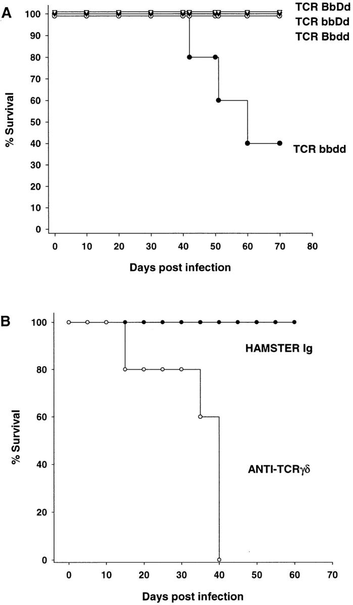
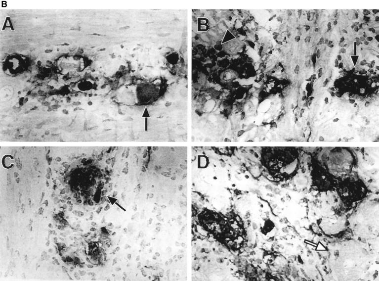
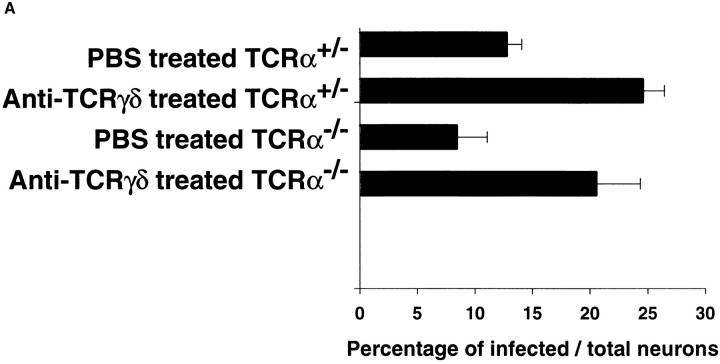
Increased numbers of T cell receptor (TCR)-gamma/delta cells have been observed in animal models of influenza and sendai virus infections, as well as in patients infected with human immunodeficiency virus and herpes simplex virus type 1 (HSV-1). However, a direct role for TCR-gamma/delta cells in protective immunity for pathogenic viral infection has not been demonstrated. To define the role of TCR-gamma/delta cells in anti-HSV-1 immunity, TCR-alpha-/- mice treated with anti- TCR-gamma/delta monoclonal antibodies or TCR-gamma/delta x TCR-alpha/beta double-deficient mice were infected with HSV-1 by footpad or ocular routes of infection. In both models of HSV-1 infection, TCR-gamma/delta cells limited severe HSV-1-induced epithelial lesions and greatly reduced mortality by preventing the development of lethal viral encephalitis. The observed protection resulted from TCR-gamma/delta cell-mediated arrest of both viral replication and neurovirulence. The demonstration that TCR-gamma/delta cells play an important protective role in murine HSV-1 infections supports their potential contribution to the immune responses in human HSV-1 infection. Thus, this study demonstrates that TCR-gamma/delta cells may play an important regulatory role in human HSV-1 infections.


Figures

Figure 1
Figure 1
TCR-γ/δ cells regulate HSV-1 infection. (A) TCR null mice were infected with HSV-1 in the hind footpad and were monitored for survival. TCR-β+/−+/− mice, open triangles; TCR-β+/−−/− mice, closed triangles; TCR-β−/−+/− mice, open circles; and TCR-β−/−/ δ−/− mice, closed circles. All groups of mice contained at least three animals, and this plot is representative of two separate experiments. (B) TCR-α−/− mice were infected with HSV-1 in the cornea and were monitored for survival. Control hamster Ig-treated TCR-α−/− mice, closed circles; anti– TCR-γ/δ mAb–treated TCR-α−/− mice, open circles. Both groups of mice contained five animals and this plot is representative of three separate experiments.
Figure 2
Figure 2
TCR-α−/− mice recover from HSV-1 infection and heal their lesions. Elimination of viral-induced vesicles and scabbing as well as initiation of new hair follicles is evident in the PBS-treated TCR-α−/− mouse on the left. In contrast, the anti–TCR-γ/δ mAb–treated TCRα−/− mouse on the right has continued vesicle formation and scabbing that covers the eye. Photos were taken at day 37 after infection from the same experiment.
Figure 3
Figure 3
The dynamics of HSV-1 replication is different in TCR-α+/− and TCR-α−/− mice. (A) Quantitation of immunohistochemical staining of HSV-1 viral antigens in the trigeminal ganglia at day 6 after infection. Two mice per group and three representative sections of each ganglia (six sections per group) were prepared for immunohistochemical staining. An average of 1,200 neurons were counted for each group. Data are recorded as the percent of neurons that exhibited specific HSV-1 staining. The differences between the day 6 after infection control and anti–TCR-γ/δ mAb–treated TCR-α+/− mice, or between the control and anti–TCR-γ/δ mAb–treated TCR-α−/− mice, were statistically significant (P <0.05). (B) Photomicrographs depicting immunohistochemical staining of HSV-1 antigens in the trigeminal ganglion. Trigeminal ganglia were obtained 6 d after corneal infection from TCRα+/− mice (A and B) and TCR-α−/− mice (C and D). Mice received control (A and C) or anti–TCR-γ/δ mAb (B and D) treatments. Infected neurons, black arrows; uninfected neurons, white arrows; a cluster of infected inflammatory cells, black arrowhead in B. Original magnification: 100×.
Figure 3
Figure 3
The dynamics of HSV-1 replication is different in TCR-α+/− and TCR-α−/− mice. (A) Quantitation of immunohistochemical staining of HSV-1 viral antigens in the trigeminal ganglia at day 6 after infection. Two mice per group and three representative sections of each ganglia (six sections per group) were prepared for immunohistochemical staining. An average of 1,200 neurons were counted for each group. Data are recorded as the percent of neurons that exhibited specific HSV-1 staining. The differences between the day 6 after infection control and anti–TCR-γ/δ mAb–treated TCR-α+/− mice, or between the control and anti–TCR-γ/δ mAb–treated TCR-α−/− mice, were statistically significant (P <0.05). (B) Photomicrographs depicting immunohistochemical staining of HSV-1 antigens in the trigeminal ganglion. Trigeminal ganglia were obtained 6 d after corneal infection from TCRα+/− mice (A and B) and TCR-α−/− mice (C and D). Mice received control (A and C) or anti–TCR-γ/δ mAb (B and D) treatments. Infected neurons, black arrows; uninfected neurons, white arrows; a cluster of infected inflammatory cells, black arrowhead in B. Original magnification: 100×.
Figure 4
Figure 4
TCR-γ/δ cells isolated from TCR-α−/− mice are specific for HSV-1 gI. Expanded TCR-γ/δ cells were stimulated with immobilized gIIg or control fusion CTLA4Ig and IFN-γ production was measured.

References

    1. Roizman, B., and A.E. Sears. 1990. Herpes simplex viruses and their replication. In Virology. B.N. Fields and D.M. Knipe, editors. Raven Press, Ltd., New York. 1795–1842.
    1. Whitley, R.J. 1990. Herpes simplex viruses. In Virology. B.N. Fields and D.M. Knipe, editors. Raven Press, Ltd., New York. 1843–1887.
    1. Schmid DS, Rouse BT. The role of T cell immunity in control of herpes simplex virus. Curr Top Microbiol Immunol. 1992;179:57–74. - PubMed
    1. Bluestone JA, Khattri R, Sciammas R, Sperling AI. TCR γδ cells: a specialized T-cell subset in the immune system. Annu Rev Cell Dev Biol. 1995;11:307–353. - PubMed
    1. Haas W, Pereira P, Tonegawa S. Gamma/delta cells. Annu Rev Immunol. 1993;11:637–685. - PubMed

Publication types

Substances