. 1997 Jun 15;17(12):4536-44.
doi: 10.1523/JNEUROSCI.17-12-04536.1997.

Abnormal synaptic plasticity in the striatum of mice lacking dopamine D2 receptors

Affiliations

Abnormal synaptic plasticity in the striatum of mice lacking dopamine D2 receptors

P Calabresi et al. J Neurosci. .

Abstract

Dopamine D2 receptors (D2Rs) are of crucial importance in the striatal processing of motor information received from the cortex. Disruption of the D2R gene function in mice results in a severe locomotor impairment. This phenotype has analogies with Parkinson's disease symptoms. D2R-null mice were used to investigate the role of this receptor in the generation of striatal synaptic plasticity. Tetanic stimulation of corticostriatal fibers produced long-term depression (LTD) of EPSPs in slices from wild-type (WT) mice. Strikingly, recordings from D2R-null mice showed the converse: long-term potentiation (LTP). This LTP, unlike LTD, was blocked by an NMDA receptor antagonist. In magnesium-free medium, LTP was also revealed in WT mice and found to be enhanced by L-sulpiride, a D2R antagonist, whereas it was reversed into LTD by LY 17555, a D2R agonist. In D2R-null mice this modulation was lost. Thus, our study indicates that D2Rs play a key role in mechanisms underlying the direction of long-term changes in synaptic efficacy in the striatum. It also shows that an imbalance between D2R and NMDA receptor activity induces altered synaptic plasticity at corticostriatal synapses. This abnormal synaptic plasticity might cause the movement disorders observed in Parkinson's disease.


Figures

Fig. 1.
Fig. 1.
In vitro intrinsic membrane properties of striatal neurons and pharmacological characteristics of corticostriatal synaptic potentials are similar for both WT (D2+/+) and D2R-null mice (D2−/−). A, The graph shows the current–voltage relationship obtained from two striatal neurons recorded from a WT (filled circles) or a D2R-null mouse (open circles). Plots were obtained from voltage-clamp experiments, holding the cells at −85 mV and applying positive and negative steps (0.5–3.0 sec duration).Right, Injection of a positive current pulse (0.9 nA) evoked a tonic firing discharge in neurons recorded from either a WT (a) or a D2R-null animal (b). In both experiments the resting membrane potential (RMP; dotted line) was −85 mV. B, The graph shows the pharmacology of the cortically evoked EPSPs recorded in WT (filled circles; n = 6) or in D2R-null mice (open circles; n = 6), either in control medium or in the absence of external magnesium.Bars indicate the time of application of APV (50 μm), CNQX (10 μm), and magnesium-free medium. Right, EPSPs recorded from single experiments in WT (a, c) or in D2R-deficient (b, d) slices, in the presence (a, b) or absence (c, d) of external magnesium. Note that APV (50 μm) reduced the EPSPs only in magnesium-free medium. RMPs were −85 mV (a) and −85 mV (b). In this figure and in the following ones arrows indicate the artifact of the single synaptic stimulation.
Fig. 2.
Fig. 2.
Tetanic stimulation of corticostriatal fibers induces LTD in slices obtained from WT mice but LTP in slices prepared from D2R-null mice. A, The graph summarizes the results from extracellular experiments, measuring the field potential amplitude, performed by using either WT (filled circles; n = 11) or D2R-null brain sections (open circles; n = 12). In this figure and in the following ones the tetanus was delivered at time 0. The bottom part of the figure shows traces from two single extracellular experiments performed with WT (a, b) or D2R-null (c, d) brain slices.B, The graph represents the results on the EPSP amplitude obtained from intracellular experiments (WT,n = 11; D2R-null, n = 9). Traces of EPSPs recorded before and after the tetanus from WT (a, b) or D2R-null (c, d) brain sections are represented in the bottom part of the figure. RMPs were −84 mV (a, b) and −85 mV (2c, d).
Fig. 3.
Fig. 3.
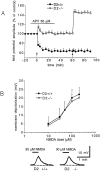
Effects of APV on synaptic plasticity and postsynaptic action of NMDA in neurons recorded from WT and D2R-null slices. A, APV (50 μm) reversibly prevented the induction of LTP in D2R-null mice (open circles; n = 13) but not the formation of LTD in WT animals (filled circles;n = 9). The bar shows the period of application of APV. Arrows indicate when the tetanic stimulation was delivered. B, The graph shows the dose–response curve for NMDA-induced membrane depolarization obtained from WT (filled circles; n = 5) and D2R-null (open circles; n = 6) slices. NMDA was bath-applied (20–30 sec) in the presence of 1 μm tetrodotoxin. The bottom part of the figure shows membrane depolarizations obtained from a WT slice (left) and from a D2R-null slice (right) after bath application of NMDA. In both cases the RMP was −85 mV.
Fig. 4.
Fig. 4.
Effects of SCH 23390 and l-sulpiride on synaptic plasticity recorded in normal medium. A, Intracellular experiments show that the pretreatment of the slices with 3 μm SCH 23390 prevented the formation of LTD in WT slices (filled circles; n = 4), but it did not affect LTP in D2R-null slices (open circles; n = 5). B, Extracellular experiments from WT slices (filled circles; n = 8) show that acute blockade of D2Rs by 1 μml-sulpiride reversibly prevented the induction of LTD but failed to cause LTP. Arrowsindicate when the tetanic stimulation was delivered. C, The traces represent field potentials recorded from WT slices before (a) and 20 min after (b) the tetanus in the presence of l-sulpiride. Traces inc and d show field potentials recorded after the washout of l-sulpiride, respectively, before and 20 min after the second tetanic stimulation.
Fig. 5.
Fig. 5.
Effects of l-sulpiride and LY 17555 on LTP recorded in magnesium-free medium from WT and D2R-null slices.A, The graph shows the long-term effects of tetanic stimulation recorded intracellularly in WT slices in the absence of external magnesium (filled circles;n = 11), in the absence of magnesium plus 1 μml-sulpiride (filled triangles; n = 7), and in magnesium-free medium plus 3 μm LY 17555 (filled squares; n = 7). Traces on the right represent EPSP recorded from WT slices in magnesium-free solution (a), in magnesium-free medium plus l-sulpiride (b), and in the absence of magnesium plus LY 17555 (c). B, The graph shows the LTP recorded in D2R-null slices in the absence of external magnesium (open circles; n = 10), in the absence of magnesium plus 1 μml-sulpiride (open triangles;n = 8), and in magnesium-free medium plus 3 μm LY 17555 (open squares;n = 9). Traces on theright represent EPSP recorded from D2R-null slices in magnesium-free solution (a), in magnesium-free medium plus l-sulpiride (b), and in the absence of magnesium plus LY 17555 (c). Time and voltage calibrations apply for both A andB.

References

    1. Albin RL, Young AB, Penney JB. The functional anatomy of basal ganglia disorders. Trends Neurosci. 1989;12:366–375. - PubMed
    1. Alexander GE, Crutcher MD. Functional architecture of basal ganglia circuits: neural substrates of parallel processing. Trends Neurosci. 1990;13:266–271. - PubMed
    1. Alexander GE, Crutcher MD, DeLong MR. Basal ganglia-thalamocortical circuits: parallel substrates for motor, oculomotor, prefrontal and limbic functions. Prog Brain Res. 1990;85:119–146. - PubMed
    1. Apicella P, Ljungberg T, Scarnati E, Schultz W. Responses to reward in monkey dorsal and ventral striatum. Exp Brain Res. 1991;85:491–500. - PubMed
    1. Artola A, Hensch T, Singer W. Calcium-induced long-term depression in the visual cortex of the rat in vitro. J Neurophysiol. 1996;76:984–994. - PubMed

Publication types

MeSH terms