. 1997 Jun 15;17(12):4662-71.
doi: 10.1523/JNEUROSCI.17-12-04662.1997.

Heterozygous peripheral myelin protein 22-deficient mice are affected by a progressive demyelinating tomaculous neuropathy

Affiliations

Heterozygous peripheral myelin protein 22-deficient mice are affected by a progressive demyelinating tomaculous neuropathy

K Adlkofer et al. J Neurosci. .

Abstract

Hereditary neuropathy with liability to pressure palsy (HNPP) is associated with a heterozygous 1.5 megabase deletion on chromosome 17 that includes the peripheral myelin protein (PMP) gene PMP22. We show that heterozygous PMP22 knock-out mice, which carry only one functional pmp22 allele and thus genetically mimic HNPP closely, display similar morphological and electrophysiological features as observed in HNPP nerves. As reported previously, focal hypermyelinating structures called tomacula, the pathological hallmarks of HNPP, develop progressively in young PMP22(+/0) mice. By following the fate of tomacula during aging, we demonstrate now that these mutant animals are also interesting models for examining HNPP disease mechanisms. Subtle electrophysiological abnormalities are detected in PMP22(+/0) mice >1 year old, and a significant number of abnormally swollen and degenerating tomacula are present. Thinly myelinated axons and supernumerary Schwann cells forming onion bulbs as fingerprints of repeated cycles of demyelination and remyelination are also encountered frequently. Quantitative analyses using electron microscopy on cross sections and light microscopy on single teased nerve fibers suggest that tomacula are intrinsically unstable structures that are prone to degeneration; however, the severity of morphological and electrophysiological abnormalities in PMP22(+/0) mice is variable. These combined findings are reminiscent of the disease progression in HNPP and offer a possible explanation about why some HNPP patients develop a chronic motor and sensory neuropathy later in life that resembles demyelinating forms of Charcot-Marie-Tooth disease by both morphological and clinical criteria.


Figures

Fig. 1.
Fig. 1.
Alkaline toluidine blue staining of quadriceps nerve cross sections of wild-type and PMP22+/0 mice of different ages. Two micrometer cross sections of the quadriceps nerve show no myelin abnormalities in 5-, 10-, and 15-month-old control animals (a, c, e). In PMP22+/0 littermates, myelinated fibers with abnormal thick myelin sheaths are visible in transverse sections of quadriceps nerves (arrowheads) (b, d, f). In 15-month-old mutants, onion bulb formation is observed (arrows) (f). Scale bar, 25 μm.
Fig. 2.
Fig. 2.
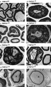
Ultrastructure of abnormal myelin in PMP22+/0 mice. Cross sections from the quadriceps nerve of a wild-type mouse (a), a 10-month-old PMP22+/0 mouse showing hypermyelination attributable to excessive wrapping of the myelin sheath (b), a 10-month-old PMP22+/0 animal illustrating the invagination of the myelin sheath as a potential start of focal hypermyelination (c), a 5-month-old PMP22+/0mouse showing intermyelin infolds forming a hypermyelin structure (d), a 10-month-old PMP22+/0 mouse showing a hypermyelin structure with displaced axon (e), a 10-month-old PMP22+/0 mouse showing degenerating hypermyelin (f), and a 15-month-old PMP22+/0 mouse showing thinly myelinated axons with concentric Schwann cell processes (g,arrowheads) and basal laminae (h,arrow) forming onion bulbs. Axons (A), compact myelin (m), and degenerating myelin (d) are marked. Scale bar, 2.5 μm.
Fig. 3.
Fig. 3.
Quantitation of prominent infoldings of the myelin sheath in 10-month-old PMP22+/0 mice. Significant numbers of myelin infolds that are extended to more than half of the axon diameter are observed on cross sections of quadriceps nerves of 10-month-old PMP22+/0 mice (n = 5). In contrast, such prominent structures were not observed in wild-type littermates (n = 2; **p ≤ 0.01; one-sided U test).
Fig. 4.
Fig. 4.
Quantitation of myelin abnormalities of PMP22+/0 mice as deduced from electron microscopy analysis. Unaffected myelinated axon–Schwann cell units are significantly reduced in 5-month-old PMP22+/0 mice, whereas hypermyelin structures are frequent. Degenerating tomacula and onion bulb formation were observed only rarely at this age. In 10-month-old PMP22+/0 mice, degenerating tomacula and onion bulb formation occurred but did not reach statistical significance; however, all three myelin abnormalities were significantly increased in 15-month-old PMP22+/0 mice in comparison with wild-type littermates (*p ≤ 0.05; one-sided Utest).
Fig. 5.
Fig. 5.
Age-related progression of myelin abnormalities in PMP22+/0 mice deduced from electron microscopy analysis. The number of hypermyelin structures does not change significantly during aging of PMP22+/0 mice, whereas degenerating hypermyelin and onion bulb formation were significantly increased in 15-month-old PMP22+/0 mice in comparison with 5-month-old mutant mice (**p ≤ 0.01; one-sidedU test).
Fig. 6.
Fig. 6.
Morphology of teased nerve fibers from wild-type and PMP22+/0 animals of different ages. Quadriceps nerve fibers in control animals (a). Para- and internodal tomacula in 5-month-old PMP22+/0 animals (b, c, large arrowheads). In 10-month-old mutant animals, enlarged tomacula presumably undergoing degeneration are observed (d, large arrow). Demyelinated internodal segments are seen in 10-month-old PMP22+/0 mice (e), but their number is increased significantly in 15-month-old mutants (f, arrowsindicate cell nuclei). Nodes of Ranvier are marked with small arrowheads (b–f). Scale bars (shown in f): a, c, d, f, 50 μm; (shown in e): b, e, 50 μm.
Fig. 7.
Fig. 7.
Irregular cytoplasmic inclusions in PMP22+/0 mice. By teased nerve fiber analysis, Schmidt-Lanterman incisures are regularly arranged in wild-type mice (a, c), whereas an unusual distribution of cytoplasmic channels is observed in mutant mice (b, d). Representative fibers are shown and phalloidin staining was used to visualize the distribution of cytoplasm (c, d). Schmidt-Lanterman incisures are marked with a star. Scale bar, 25 μm.
Fig. 8.
Fig. 8.
Progression of myelin abnormalities in affected internodal segments in teased nerve fibers of PMP22+/0mice. The number of tomaculous internodes decreases significantly with age. In contrast, an increase in degenerating tomacula is observed in 10- and 15-month-old PMP22+/0 mice but reaches significance only at 15 months. The observed increase in onion bulb formation is significant at 15 months (*p ≤ 0.05, **p ≤ 0.01; one-sided Utest).

References

    1. Adlkofer K, Martini R, Aguzzi A, Zielasek J, Toyka KV, Suter U. Hypermyelination and demyelinating peripheral neuropathy in pmp22-deficient mice. Nature Genet. 1995;11:274–280. - PubMed
    1. Amato AA, Gronseth GS, Callerame KJ, Kagan-Hallet KS, Bryan WW, Barohn RJ. Tomaculous neuropathy: a clinical and electrophysiological study in patients with and without 1.5-Mb deletions in chromosome 17p11.2. Muscle Nerve. 1996;19:16–22. - PubMed
    1. Anzini P, Neuberg DH-H, Schachner M, Nelles E, Willecke K, Zielasek J, Toyka K, Suter U, Martini R. Structural abnormalitites and deficient maintenance of peripheral nerve myelin in mice lacking the gap junction protein connexin 32. J Neurosci. 1997;17:4545–4551. - PMC - PubMed
    1. Chance PF, Alderson MK, Leppig KA, Lensch MW, Matsunami N, Smith B, Swanson PD, Odelberg SJ, Disteche CM, Bird TD. DNA deletion associated with hereditary neuropathy with liability to pressure palsies. Cell. 1993;72:143–151. - PubMed
    1. Davies DM. Recurrent peripheral nerve palsies in a family. Lancet. 1954;2:266–268. - PubMed

Publication types

MeSH terms