. 1997 Jun 15;17(12):4700-10.
doi: 10.1523/JNEUROSCI.17-12-04700.1997.

The shaking-B2 mutation disrupts electrical synapses in a flight circuit in adult Drosophila

Affiliations

The shaking-B2 mutation disrupts electrical synapses in a flight circuit in adult Drosophila

J R Trimarchi et al. J Neurosci. .

Abstract

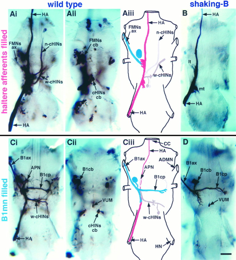
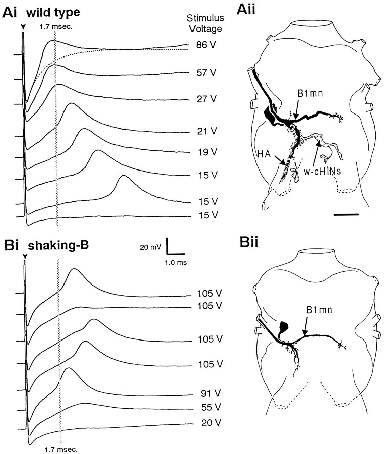
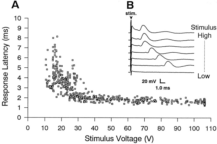
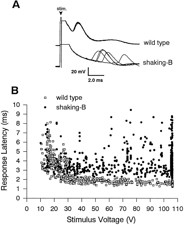
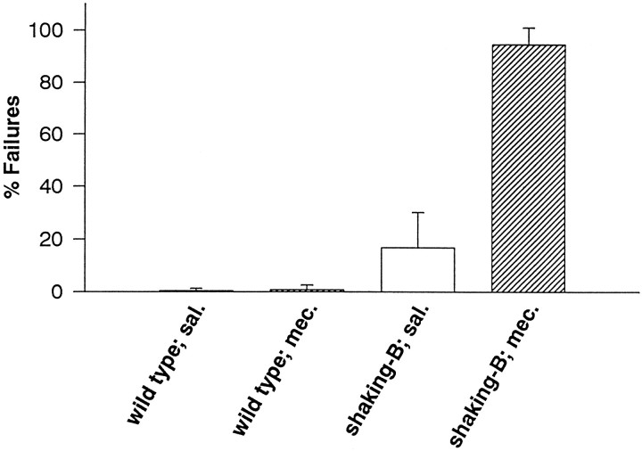
The shaking-B2 mutation was used to analyze synapses between haltere afferents and a flight motoneuron in adult Drosophila. We show that the electrical synapses among many neurons in the flight circuit are disrupted in shaking-B2 flies, suggesting that shaking-B expression is required for electrical synapses throughout the nervous system. In wild-type flies haltere afferents are dye-coupled to the first basalar motoneuron, and stimulation of these afferents evokes electromyograms from the first basalar muscle with short latencies. In shaking-B2 flies dye coupling between haltere afferents and the motoneuron is abolished, and afferent stimulation evokes electromyograms at abnormally long latencies. Intracellular recordings from the motoneuron confirm that the site of the defect in shaking-B2 flies is at the synapses between haltere afferents and the flight motoneuron. The nicotinic cholinergic antagonist mecamylamine blocks the haltere-to-flight motoneuron synapses in shaking-B2 flies but does not block those synapses in wild-type flies. Together, these results show that the haltere-to-flight motoneuron synapses comprise an electrical component that requires shaking-B and a chemical component that is likely to be cholinergic.


Figures

Fig. 1.
Fig. 1.
Dye coupling is eliminated inshaking-B2 flies. A, Anterograde staining of haltere afferents in a wild-type fly. Shown is a montage of dorsal projections (Ai) and ventral cell bodies (Aii). Aiii, Schematic derived from Ai and Aii illustrating the prominent neurons dye-coupled to haltere afferents (see Tables 1, 2 for frequency of occurrence). Nerve trunks are labeled inCiii. Stained haltere afferents (HA;red) are visible in the haltere nerve and extend through the dorsal region of the ganglia before exiting through the cervical connective. Dye passes from haltere afferents to flight motoneurons identified by staining of axons (FMNs ax;blue) in the ADMN and large ventral cell bodies (FMNs cb; blue). Dye also passes to several large interneurons [neck (n-cHINs) and wing contralaterally projecting haltere interneurons (w-cHINs; gray)] with cell bodies (cHINs cb; gray) located in the ventroposterior rind of the mesothoracic leg neuromere.B, Anterograde staining of haltere afferents in ashaking-B2 fly. Dye did not pass from haltere afferents to any other neurons. Stained haltere afferents (HA) are visible in haltere nerve and extend through the dorsal region of the ganglia, forming a medial tuft (mt) and lateral tuft (lt) of arbors before exiting through the cervical connective. C, Retrograde staining of the B1mn in a wild-type fly. Shown is a montage of dorsal projections (Ci) and ventral cell bodies (Cii).Ciii, Schematic derived from Ci andCii illustrating the prominent neurons dye-coupled to B1mn (see Tables 1, 2 for frequency of occurrence). The B1mn axon (B1ax; blue) is visible in the ADMN, and the contralateral process (B1cp; blue) crosses the midline through the mesothoracic decussation. B1mn has a large ventrally located cell body (B1cb). Dye passes from B1mn (blue) to haltere afferents (HA; red). Neurobiotin appears to travel preferentially in the retrograde direction; thus haltere afferent projections that extend anteriorly through the cervical connective were stained only faintly. Dye also passed to several interneurons (w-cHINs; gray) with cell bodies (cHINs cb; gray) located in the ventroposterior rind of the mesothoracic leg neuromere. Note that the w-cHINs dye-coupled to B1mn appear to be contralateral homologs of those coupled to haltere afferents. Several anteriorly projecting neurons (APNs;gray) also were dye-coupled to B1mn. D, Retrograde staining of B1mn in a shaking-B2fly. Dye did not pass from B1mn to any other neurons. The B1mn axon (B1ax) is visible in the ADMN, and the contralateral process (B1cp) crosses the midline through the mesothoracic decussation. The ventral unpaired median cell (VUM) that also innervates B1 is visible inCii and D. Scale bar, 20 μm.
Fig. 4.
Fig. 4.
Intracellular recordings from B1mn.Ai, Action potentials recorded intracellularly from B1mn evoked by haltere afferent stimulation in a wild-type fly. Low-voltage stimuli evoke action potentials that occur at long latencies. As the stimulus intensity is increased, the latency from the stimulus to the action potential decreases. Strong stimuli evoke action potentials that occur at extremely short latencies (<1.7 msec), resulting in the peak of the action potential being partially obscured by the stimulus artifact. The dotted line in the top trace is the stimulus artifact when the electrode was adjacent to, but not in, the cell and can be used as a reference to illustrate that the action potential is obscured partially by the stimulus artifact. The arrowhead marks the stimulus artifact. Thegray vertical line is at 1.7 msec and denotes the average latency at which haltere afferent stimulation evokes short-latency EMGs from the B1 muscle in wild type (see Fig. 2).Aii, Camera lucida drawing of an intracellularly stained B1mn from a wild-type fly (dorsal view). Neurobiotin was injected intracellularly into B1mn and passed to haltere afferents and w-cHINs. Scale bar, 20 μm. Bi, Action potentials recorded intracellularly from B1mn evoked by haltere afferent stimulation in ashaking-B2 fly. At all stimulus voltages action potentials were evoked with variable and abnormally long latencies (>1.7 msec). Unlike in wild-type flies, action potentials inshaking-B2 flies were never obscured by the stimulus artifact. Occasionally, strong stimuli did not evoke action potentials in shaking-B2 flies (second trace from the top).Bii, Camera lucida drawing of an intracellularly stained B1mn from a shaking-B2 fly (dorsal view). Neurobiotin was injected intracellularly into B1mn and did not pass to any other neurons. Scale bar is the same as in Ai.
Fig. 2.
Fig. 2.
In wild-type flies stimulation of haltere afferents evokes EMGs from B1. A, Summary of B1 EMG latencies evoked by a range of stimulus intensities (n = 6 flies). Low-voltage stimuli (<30 V) evoke B1 EMGs at long and variable latencies (2.1–8.5 msec), consistent with the activation of B1mn via a polysynaptic pathway. Stronger stimuli evoke B1 EMGs at a constant short latency of, on average, 1.7 msec, consistent with the activation of B1mn via monosynaptic electrical synapses. All six flies displayed similar response profiles, exhibiting B1 EMGs at both long and short latencies. B, Sample B1 EMGs evoked by an ascending series of stimulus intensities applied to haltere afferents. The bottom trace was evoked by the weakest stimulus intensity, and as the stimulus intensity was increased, the latency from the stimulus to the evoked EMG from B1 decreased. The arrowhead marks the stimulus artifact. Response latencies were measured from the onset of the stimulus to the onset of the evoked EMG.
Fig. 3.
Fig. 3.
In shaking-B2flies stimulation of haltere afferents evokes EMGs from B1 with abnormally long latencies. A, B1 EMGs evoked by haltere afferent stimulation at 80 V. The five B1 EMGs evoked consecutively from a wild-type fly occur at similar latencies (∼1.7 msec). In contrast, the five B1 EMGs evoked consecutively from ashaking-B2 fly occur at longer (>2.1 msec) and more variable latencies. The arrowhead marks the stimulus artifact. B, Summary of B1 EMGs latencies evoked by a range of stimulus intensities (n = 6shaking-B2 flies, 6 wild-type flies). The wild-type data (□) have been replotted from Figure 2 and serve as a reference. Low-voltage stimuli (<50 V) evoke B1 EMGs from bothshaking-B2 (•) and wild-type flies that occur at similar long and variable latencies (2.1–9.5 msec). By contrast, stimuli (>50 V) that evoke B1 EMGs at latencies <2.1 msec in all wildtype flies tested (n = 6) evoke B1 EMGs at latencies >2.1 msec in allshaking-B2 flies tested (n = 6). Importantly, many of the strong stimuli evoke EMGs from shaking-B2 flies that occur at latencies 0.5–2.2 msec longer than 1.7 msec (see Discussion). The vertical cluster of shaking-B2 data points at the far right of the graph result from stimuli of 105 V that evoked B1 EMGs occurring at a wide range of latencies (2.1–9.5 msec). A similar number of data points were gathered from wild-type flies but exhibited remarkably constant latencies (<2.1 msec), and thus the plotted points overlap (also see Fig. 2).
Fig. 5.
Fig. 5.
Mecamylamine blocks B1 EMGs evoked by haltere afferent stimulation in shaking-B2 flies, but not wild-type flies. The percentage of stimuli that failed to evoke EMGs in shaking-B2 flies was increased dramatically by the presence of mecamylamine (17% in saline; 95% in mecamylamine). The percentage of stimuli that failed to evoke EMGs in wild-type flies was not increased by the presence of mecamylamine (<1% in saline; <1% in mecamylamine).

References

    1. Albert JL, Lingle CJ. Activation of nicotinic acetylcholine receptors on cultured Drosophila and other insect neurons. J Physiol (Lond) 1993;463:605–630. - PMC - PubMed
    1. Baird DH, Schalet AP, Wyman RJ. The Passover locus in Drosophila melanogaster: complex complementation and different effects on the giant fiber neural pathway. Genetics. 1990;126:1045–1059. - PMC - PubMed
    1. Baird DH, Koto M, Wyman RJ. Dendritic reduction in Passover, a Drosophila mutant with defective giant fiber neuronal pathway. J Neurobiol. 1993;24:971–984. - PubMed
    1. Balakrishnan R, Rodrigues V. The shaker and shaking-B genes specify elements in the processing of gustatory information in Drosophila melanogaster. J Exp Biol. 1991;157:161–181. - PubMed
    1. Bennett MVL. Electrical transmission: a functional analysis and comparison to chemical transmission. In: Brookhart JM, Mountcastle VB, editors. Handbook of physiology: the nervous system. American Physiological Society; Bethesda, MD: 1977. pp. 357–416.

Publication types

LinkOut - more resources