. 1997 Jun 15;17(12):4764-84.
doi: 10.1523/JNEUROSCI.17-12-04764.1997.

Glutamatergic enteric neurons

Affiliations

Glutamatergic enteric neurons

M T Liu et al. J Neurosci. .

Abstract

We tested the hypothesis that glutamate, the major excitatory neurotransmitter of the CNS, is also an excitatory neurotransmitter in the enteric nervous system (ENS). Glutamate immunoreactivity was found in cholinergic enteric neurons, many of which were identified as sensory by their co-storage of substance P and/or calbindin. Glutamate immunoreactivity was concentrated in terminal varicosities with a majority of small clear synaptic vesicles. The immunoreactivities of both AMPA and NMDA receptor subunits were also detected on neurons in both submucosal and myenteric plexuses. The immunoreactivity of the EAAC1 neuronal glutamate transporter was widespread in both plexuses. Glutamate evoked depolarizing responses in myenteric neurons that had fast and slow components. The fast component was mimicked by AMPA, and the slow component was mimicked by NMDA. The fast component and the response to AMPA mimicked fast EPSPs evoked in 2/AH neurons; moreover, fast EPSPs as well as fast glutamate and AMPA responses were blocked by selective AMPA antagonists and potentiated by the glutamate uptake inhibitor L-(-)-threo-3-hydroxyaspartic acid. These observations demonstrate, for the first time, the presence of glutamatergic neurons and glutamate-mediated neurotransmission in the ENS.


Figures
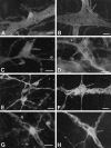
Fig. 1.
Fig. 1.
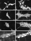
Glutamate immunoreactivity in the guinea pig and rat small intestine. A–D, Guinea pig ileum. A subset of neurons in the myenteric (A) and submucosal (C) plexus are glutamate-immunoreactive (arrow). Varicose (arrowhead) and smooth (arrow) glutamate-immunoreactive nerve fibers are found in myenteric (B) and submucosal (D) ganglia. Glutaraldehyde-induced background autofluorescence is present. E, F, Rat ileum. Glutamate-immunoreactive neurons (arrow) and nerve fibers (arrowhead) are numerous in submucosal (E) and myenteric (F) ganglia. G, H, Guinea pig ileum. Glutamate immunoreactivity (G) is expressed by all substance P-immunoreactive neurons (H) in the submucosal plexus (arrow). Scale bar, 30 μm.
Fig. 2.
Fig. 2.
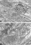
Glutamate-immunoreactive varicosities can be detected by electron microscopy in myenteric ganglia. A, Small, clear, round synaptic vesicles (arrow) are present in all glutamate-immunoreactive nerve terminals (t). The black dots(arrowhead) are gold particles (10 nm), which identify the axon as containing high levels of glutamate. A second terminal (*) also contains clear, round synaptic vesicles (arrow) but few gold particles. Note that there is some diffusion of the amino acid. B, Varicosities in which large, dense-cored vesicles (arrow) predominate are not labeled by antibodies to glutamate. Scale bar, 0.5 μm.
Fig. 3.
Fig. 3.
Differential distribution of AMPA receptor subunit immunoreactivity in the guinea pig small intestine. A, B, GluR1 immunoreactivity. A subset of neurons in the submucosal (A) and myenteric (B) plexus express GluR1 immunoreactivity. Immunoreactivity is concentrated in the cytoplasm. C, D, GluR2/3 immunoreactivity. The majority of neurons in the submucosal plexus (C) express GluR2/3 immunoreactivity. A subset of neurons in the myenteric plexus (D) expresses GluR2/3. These cells are small in size (arrow). E, F, GluR4 immunoreactivity. A subset of neurons in the submucosal (E) and myenteric (F) plexus express GluR4 immunoreactivity. Immunoreactivity is concentrated in the cytoplasm; however, proximal dendritic processes are also stained (arrow). G, H, GluR1 immunoreactivity (G) is expressed by the majority of calbindin-immunoreactive myenteric neurons (H, arrow). Scale bar, 30 μm.
Fig. 4.
Fig. 4.
NMDA receptor subunit immunoreactivity in the guinea pig small intestine. A, B, The majority of neurons in the submucosal (A) and myenteric (B) plexus express NR2A/B immunoreactivity. Immunoreactivity is generally diffuse within labeled cells; however, labeled puncta are also observed (arrow).
Fig. 5.
Fig. 5.
Immunostaining of Western blots of guinea pig LMMP with antibodies to GluR1 (lane 1), GluR2/3 (lane 2), GluR4 (lane 3), NR1 (lane 4), NRA/B (lane 5), and EAAC1 (lane 6). Forty micrograms of protein were applied to each lane. Arrows indicate positions of molecular weight standards myosin, β-galactosidase, phosphorylase B, BSA, and ovalbumin. Lanes 1–6 were done on separate gels, and the positions of the bands cannot be compared directly.
Fig. 6.
Fig. 6.
Glutamate (Glut) depolarizes 1/S and 2/AH myenteric neurons. Fast- and slow-depolarizing responses to glutamate are observed in 1/S and 2/AH cells. The downward deflections represent the electrotonic responses to the injections of hyperpolarizing current pulses. 1, Application of glutamate (arrow) induces a transient depolarization (fast response; arrowhead) that is associated with a decrease in input resistance (reflected by a decline in the amplitude of electrotonic potentials). Note that the 2/AH cell spikes during the fast response. 2, Microejection of glutamate leads to a prolonged membrane depolarization (slow response) associated with an increase in action potential activity. 3, Application of glutamate induces a biphasic depolarizing response, consisting of an initial fast response (arrowhead) and a partial recovery of the membrane potential, followed by a slow response. The cells discharge action potentials during the fast and slow response. For 1/S cells, resting membrane potential (RMP) = −47, −42, and −49 mV; for 2/AH cells, RMP = −58, −61, and −56 mV.
Fig. 7.
Fig. 7.
Concentration dependence of glutamate-mediated depolarizations. A, Glutamate was pressure-applied (arrow) at the indicated pulse durations (in milliseconds). Responses were obtained from three different 2/AH cells (RMP = −58, −61, and −56 mV, respectively). B, Summary of concentration dependence of glutamate-mediated slow depolarizations. Data are expressed as percentages of maximum control responses (n = 6).
Fig. 8.
Fig. 8.
Responses to glutamate (Glut) in myenteric neurons are direct and not attributable to the release of another neurotransmitter. 1, Microejection of glutamate onto a 2/AH neuron causes a fast response (arrowhead, Control) that is not inhibited by a low-Ca2+/high-Mg2+-containing solution. The cell spikes repetitively in the low-Ca2+/high-Mg2+-containing solution because a Ca2+-activated K+ conductance contributes to the RMP of 2/AH cells, which become depolarized in the absence of Ca2+.2, Superfusion with TTX has no effect on glutamate-induced depolarizations recorded in a 2/AH neuron. Note that Ca2+ spikes are observed in the presence of TTX.
Fig. 9.
Fig. 9.
Glutamate-induced fast-depolarizing responses in enteric neurons are mediated by postsynaptic AMPA receptors. A, B, Fast responses of enteric neurons to AMPA can be distinguished from those to nicotine. Similar fast responses are elicited by microejection of AMPA and nicotine in 1/S neurons (Control). The fast response to AMPA is antagonized by the non-NMDA antagonist CNQX (A); however, CNQX does not affect responses to nicotine (A). The nicotinic antagonist hexamethonium does not affect responses to AMPA (B); however, it blocks responses to nicotine (B). Recordings inA and B were obtained from different neurons.
Fig. 10.
Fig. 10.
Fast responses to glutamate (Glut) are antagonized by DNQX. A, Fast responses to glutamate were recorded in a 2/AH neuron (Control). Superfusion with DNQX blocks the fast response. B, Desensitization of AMPA receptors limits AMPA-mediated fast responses. Superfusion with cyclothiazide, a selective blocker of AMPA receptor desensitization, potentiates fast responses to AMPA recorded in a 2/AH neuron.
Fig. 11.
Fig. 11.
NMDA mimics the slow response to glutamate (Glut). A, Microejection of glutamate (arrow) onto a 2/AH neuron (RMP = −57 mV) leads to a prolonged membrane depolarization (slow response) associated with spike activity. Superfusion with DNQX does not block the glutamate-mediated slow response. B, Microejection of NMDA (arrow) onto a 2/AH neuron (RMP = −58 mV) leads to a prolonged membrane depolarization associated with spike activity. The NMDA-mediated depolarization is blocked by AP5, an NMDA antagonist. C, The NMDA-mediated depolarization recorded in a 2/AH neuron (RMP = −56 mV) is blocked by the NMDA antagonist CPP. Responses to NMDA in B and C were obtained in Mg2+-free Krebs’ solution that contained glycine (10 μm). D, Glutamate evokes fast and slow responses (associated with spike activity) in a 2/AH neuron (RMP = −58 mV). Superfusion with AP5 inhibits only the slow response to glutamate.
Fig. 12.
Fig. 12.
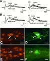
Fast synaptic transmission in 2/AH neurons is attributable to activation of AMPA receptors. A, B, Fast EPSPs in 2/AH neurons are blocked by AMPA receptor antagonists. Effects of DNQX (left) and hexamethonium (right) on fast EPSPs recorded in 2/AH (A) and 1/S (B) neurons. DNQX blocks the fast EPSP recorded in a 2/AH neuron but only slightly reduces the amplitude of the fast EPSP recorded in a 1/S neuron. In contrast, the amplitude of the fast EPSP in a 2/AH neuron is only slightly reduced by hexamethonium; however, the fast EPSP recorded in a 1/S cell is completely blocked by hexamethonium. Note that the recordings are obtained from different cells. Washindicates recovery from DNQX or hexamethonium. C–F, Glutamate-responsive enteric neurons express GluR1 immunoreactivity. Fast responses to glutamate were recorded; neurons that did or did not respond to glutamate are marked by intracellular injection of Neurobiotin. A glutamate-responsive 2/AH neuron (arrow; this neuron also displayed a glutamatergic fast EPSP) is marked by both Neurobiotin (FITC; D) and GluR1 immunoreactivity (Cy3;C). In contrast, a Neurobiotin-injected 2/AH cell that did not respond to glutamate does not contain GluR1-immunoreactivity (D, arrowhead). E, F, A Neurobiotin-injected 1/S neuron (FITC; F) that did not respond to glutamate does not express GluR1 immunoreactivity (E, arrow). Scale bar, 30 μm.
Fig. 13.
Fig. 13.
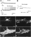
A, B, Inhibition of glutamate uptake potentiates both glutamate-mediated depolarizations and fast EPSPs in 2/AH neurons. Superfusion with THA, a specific high-affinity glutamate transport inhibitor, potentiates and prolongs the fast response to glutamate (A) and the fast EPSP recorded in a 2/AH neuron (B). C, D, Glutamate-responsive enteric neurons express the neuronal glutamate transporter EAAC1. A glutamate-responsive 2/AH neuron was marked by intracellular injection of Neurobiotin (C, arrow). The glutamate-responsive 2/AH neuron (C) is EAAC1-immunoreactive (D, arrow). Neurobiotin was visualized with avidin–FITC. EAAC1 immunoreactivity was visualized with Cy3. E, F, EAAC1 immunoreactivity (E, arrow) is expressed by all calbindin-immunoreactive neurons (F, arrow) in the myenteric plexus; however, more neurons express EAAC1 immunoreactivity (arrowhead) than calbindin. Scale bar, 30 μm.
Fig. 14.
Fig. 14.
EAAC1-immunoreactive neurons are found in the submucosal plexus. A, B, A subset of EAAC1-immunoreactive neurons (A) expresses calbindin immunoreactivity (B, arrow). C, D, EAAC1 immunoreactivity (C) is expressed by all substance P-immunoreactive neurons (D, arrow); however, not all EAAC1-immunoreactive neurons express substance P immunoreactivity (arrowhead). E, F, Confocal photomicrographs (sum of 10 optical sections, collected at 0.5 μm intervals) of a whole mount of the submucosal plexus. EAAC1-immunoreactive nerve fibers encircle intestinal crypts (E, arrow). EAAC1 immunoreactivity is found in a subset of enterochromaffin cells (F). Thearrows indicate prominent staining of the apex of the cells facing the intestinal lumen. Scale bar, 30 μm.

References

    1. Ascher P, Johnson JW. The NMDA receptor, its channel, and its modulation by glycine. In: Collingridge GL, Watkins JC, editors. The NMDA receptor. Oxford UP; Oxford: 1994. pp. 177–205.
    1. Balcar V, Johnston GAR, Twitchin B. Stereospecificity of the inhibition of l-glutamate and l-aspartate high affinity uptake in the rat brain by threo-3-hydroxyaspartate. J Neurochem. 1977;28:1145–1146. - PubMed
    1. Bertrand PP, Galligan JJ. Signal-transduction pathways causing slow synaptic excitation in guinea pig myenteric AH neurons. Am J Physiol. 1995;269:G710–G720. - PubMed
    1. Bornstein J. Local neural control of intestinal motility: nerve circuits deduced for the guinea-pig small intestine. Clin Exp Pharmacol Physiol. 1994;21:441–452. - PubMed
    1. Bornstein JC, Hendriks R, Furness JB, Trussell DC. Ramifications of the axons of AH-neurons injected with the intracellular marker biocytin in the myenteric plexus of the guinea pig small intestine. J Comp Neurol. 1991;314:437–451. - PubMed

Publication types

MeSH terms

LinkOut - more resources