. 1997 Jun 15;17(12):4839-48.
doi: 10.1523/JNEUROSCI.17-12-04839.1997.

Dual ultrastructural localization of mu-opioid receptors and NMDA-type glutamate receptors in the shell of the rat nucleus accumbens

Affiliations

Dual ultrastructural localization of mu-opioid receptors and NMDA-type glutamate receptors in the shell of the rat nucleus accumbens

K N Gracy et al. J Neurosci. .

Abstract

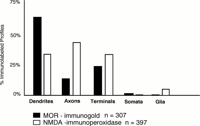
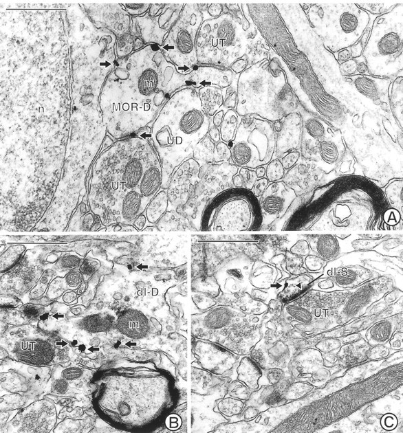
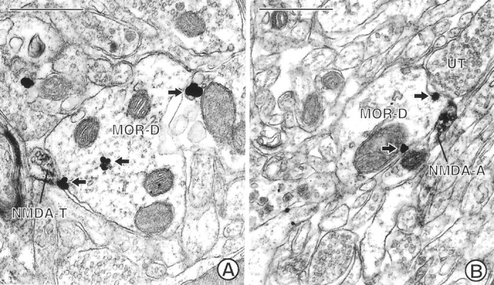
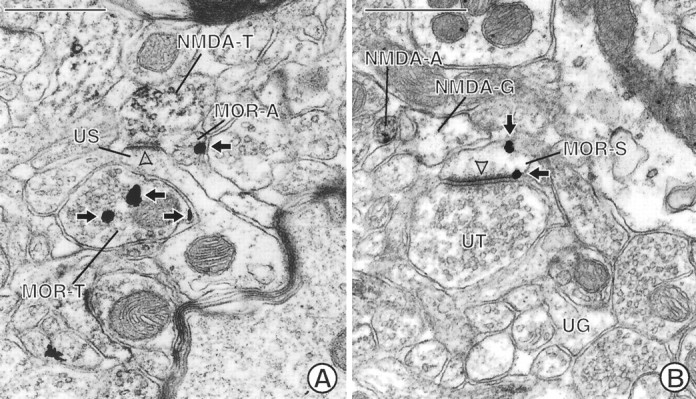
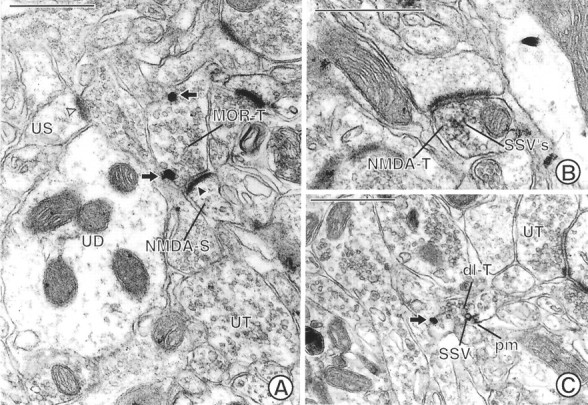
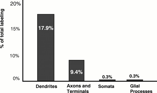
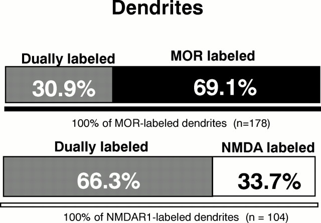
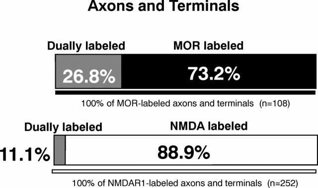
The effectiveness of NMDA antagonists in modulating the motor and motivational effects of opiates is attributed, in part, to functional associations involving NMDA receptors and micro-opioid receptors (MORs) in the shell of the nucleus accumbens (Acb). To determine the subcellular sites for potential functional interactions between opiate ligands and NMDA receptors in this region, we examined the ultrastructural localization of antipeptide antisera against MOR and the R1 subunit of the NMDA receptor in the Acb shell of the adult rat brain. MOR-like immunoreactivity (MOR-LI) was seen primarily in dendrites, whereas NMDAR1-like immunoreactivity (NMDAR1-LI) was detected more often in axon terminals forming asymmetric synapses. In these profiles, MOR labeling was localized mainly to extrasynaptic plasma membranes, whereas NMDAR1-LI was associated with both synaptic and extrasynaptic sites. Of 307 MOR-labeled processes, 17.9% of the dendrites and 9.4% of the axon terminals also contained NMDAR1-LI. In addition, 24.7% of the dendrites containing only MOR-LI were apposed to NMDAR1-labeled axons or terminals. We conclude that in the shell of the Acb, the output of single neurons can be dually modulated by (1) activation of MOR and NMDA receptors in the same dendrites or (2) combined activation of presynaptic NMDA receptors in afferents contacting dendrites containing MOR. In addition, the colocalization of MOR and NMDAR1 in certain axon terminals in the Acb suggests their dual involvement in the presynaptic release of neurotransmitters in this region.


Figures

Fig. 1.
Fig. 1.
Bar graph showing the distribution of immunogold-labeled MOR and immunoperoxidase-labeled NMDAR1 in neuronal compartments (dendrites, axons, terminals, somata) and in glial processes in the Acb. n = 307 MOR-labeled profiles and n = 397 NMDA-labeled profiles seen in 7640 μm2 area of tissue.
Fig. 2.
Fig. 2.
Electron micrographs through the Acb showing single MOR labeling and dual MOR and NMDAR1 labeling in spiny dendrites. A, A spiny dendrite (MOR-D) showing plasmalemmal immunogold labeling (arrows) for MOR is apposed to two unlabeled terminals (UT) and an unlabeled dendrite (UD). n, Nucleus; m, mitochondrion. B, A dendrite (dl-D) labeled with both immunogold (arrows) for MOR and immunoperoxidase for NMDAR1.UT, Unlabeled terminal. The electron dense peroxidase reaction product is most evident in the region above a mitochondrion (m) in the dendrite. C, A dually labeled small dendritic spine (dl-S) shows immunogold (arrow) MOR labeling along a nonsynaptic region of the plasma membrane. In contrast, immunoperoxidase for NMDAR1 is present within the asymmetric, excitatory-type contact (closed arrowhead) from an unlabeled terminal (UT). The peroxidase reaction product is also diffusely localized within the cytoplasm and the nonsynaptic regions of the plasma membrane. Scale bars, 0.5 μm.
Fig. 3.
Fig. 3.
Electron micrographs showing MOR-immunoreactive dendrites in contact with NMDAR1-labeled terminals. A, A dendrite (MOR-D) labeled with immunogold (arrows) for MOR is apposed by a small terminal (NMDA-T) labeled with immunoperoxidase for NMDAR1. B, A small preterminal axon (NMDA-A) containing immunoperoxidase labeling for NMDAR1 apposes a dendrite (MOR-D) labeled with immunogold (arrows) for MOR. The dendrite is apposed by an unlabeled terminal (UT). Scale bars, 0.5 μm.
Fig. 4.
Fig. 4.
Electron micrographs showing appositional contacts between MOR-labeled neurons and NMDAR1-labeled neuronal and glial processes. A, An unlabeled dendritic spine (US) is apposed to a terminal (MOR-T) labeled with immunogold (arrows) for MOR. The spine also receives an asymmetric, excitatory-type contact (open arrowhead) from a terminal (NMDA-T) labeled with immunoperoxidase for NMDAR1. An apposed small unmyelinated axon (MOR-A) contains immunogold labeling (arrow) along the plasma membrane. B, A glial process (NMDA-G) shows immunoperoxidase reaction product that is recognizable by the granular precipitate within the cytoplasm and along its irregularly shaped plasma membrane. The peroxidase product can be seen by comparison with an unlabeled glial process (UG) in the micrograph. The labeled glial process apposes a dendritic spine (MOR-S) containing immunogold (arrows) for MOR on nonsynaptic regions of the plasma membrane. The spine receives an asymmetric contact (open arrowhead) from an unlabeled terminal (UT). NMDA-A, NMDAR1-labeled axon. Scale bars, 0.5 μm.
Fig. 5.
Fig. 5.
Electron micrographs showing labeling for MOR and/or NMDAR1 in axon terminals in the Acb. A, A terminal (MOR-T) shows immunogold labeling (arrows) for MOR along nonsynaptic regions of the plasma membrane. This terminal forms an asymmetric synapse with a dendritic spine (NMDA-S) containing immunoperoxidase reaction product for NMDA. The immunoperoxidase reaction product is associated with the asymmetric junction (closed arrowhead), which is notably more electron dense than that seen in an unlabeled spine (US) postsynaptic density (open arrowhead). UT, Unlabeled terminal;UD, unlabeled dendrite. B, A terminal (NMDA-T) showing dense immunoperoxidase reaction product for NMDAR1 that rims the membranes of many small synaptic vesicles (SSVs) forms an asymmetric contact on a dendritic spine. C, A dually labeled axon terminal (dl-T) contains both immunogold (arrow) labeling for MOR and immunoperoxidase labeling for NMDAR1. The gold particle is located on the plasma membrane of the terminal. The peroxidase reaction product is also associated with the plasma membrane (pm) and the membrane of adjacent SSVs. UT, Unlabeled terminal. Scale bars, 0.5 μm.
Fig. 6.
Fig. 6.
Bar graph showing the relative frequencies of colocalization of MOR and NMDAR1 in neuronal (dendrites, axons, and terminals) and glial processes in the Acb. The distribution represents the 28% (86/307) of immunogold MOR-labeled profiles that contained immunoperoxidase NMDAR1 labeling from 7640 μm2 area of tissue.
Fig. 7.
Fig. 7.
Bar graphs showing proportion of single versus dual labeling in MOR- and NMDAR1-labeled dendrites in the Acb. Analysis carried out in 7640 μm2 area of tissue processed for immunogold labeling of MOR and immunoperoxidase detection of NMDAR1.
Fig. 8.
Fig. 8.
Bar graph showing proportions of single versus dual labeling in MOR- and NMDAR1-labeled axons and terminals in the Acb. Analysis carried out in 7640 μm2 area of tissue processed for immunogold labeling of MOR and immunoperoxidase detection of NMDAR1.

References

    1. Arvidsson U, Riedl M, Chakrabarti S, Lee J, Nakano A, Dado R, Loh H, Law P, Wessendorf M, Elde R. Distribution and targeting of a μ-opioid receptor (MOR1) in brain and spinal cord. J Neurosci. 1995;15:3328–3341. - PMC - PubMed
    1. Azaryan AV, Coughlin LJ, Buzas B, Clock BJ, Cox BM. Effects of chronic cocaine treatment on mu- and delta-opioid receptor mRNA levels in dopaminergically innervated brain regions. J Neurochem. 1996;66:443–448. - PubMed
    1. Ben-Eliyahu S, Marek P, Vaccarino AL, Mogil JS, Sternberg WF, Liebeskind JC. The NMDA receptor antagonist MK-801 prevents long-lasting non-associative morphine tolerance in the rat. Brain Res. 1992;575:304–308. - PubMed
    1. Bouyer JJ, Miller RJ, Pickel VM. Ultrastructural relation between cortical efferents and terminals containing enkephalin-like immunoreactivity in rat neostriatum. Regul Pept. 1984;8:105–115. - PubMed
    1. Brose N, Huntley GW, Stern BY, Sharma G, Morrison JH, Heinemann SF. Differential assembly of coexpressed glutamate receptor subunits in neurons of rat cerebral cortex. J Biol Chem. 1994;269:16780–16784. - PubMed

Publication types

Substances