

Review
doi: 10.1073/pnas.94.12.5986.
Fas-ligand: privilege and peril
Affiliations
- PMID: 9177153
- PMCID: PMC33671
- DOI: 10.1073/pnas.94.12.5986
Item in Clipboard
Review
Fas-ligand: privilege and peril
Proc Natl Acad Sci U S A.
.
No abstract available
Figures
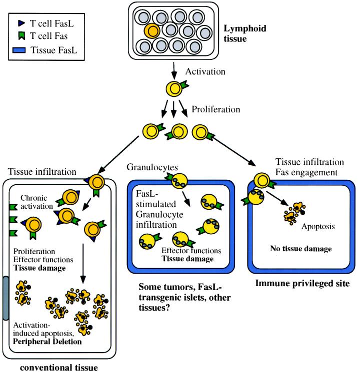
Some immunological effects of FasL. Chronically
activated T lymphocytes express both Fas and FasL, and in conventional
tissues (Left) this can result in apoptotic
death of the T cells (peripheral deletion) and induction of
apoptosis in other Fas-expressing cells. Immunologically
privileged tissues (Right) constitutively express FasL,
and infiltrating T cells and granulocytes rapidly undergo
apoptosis. Thus, the tissue is protected from any damage that
might result from an immune response. In some tissues, however
(Center), FasL induces a granulocytic infiltration,
which can damage the tissue. The conditions that favor one or the other
of these contrasting effects of tissue FasL are unknown.
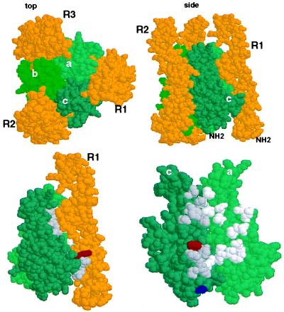
The E218G allelic substitution lies in the
receptor binding region of Fas Ligand. (Upper)
Space-filling depiction of LTα TNFR60 ligand–receptor complex from
the crystal structure derived by Banner et al. (17)
(viewed with RasMol). The three receptor (R) chains (gold) surround the
LTα subunits (green) that form the trimeric ligand. The upper left
panel (top) is viewed from the perspective of the receptor-expressing
cell with the receptor’s N terminus extending away from the reader; in
the right panel (side) the N terminus of the elongated receptor
protrudes away from the cell surface. (Lower) Location
of the T184A and E218G polymorphisms of FasL in the structure of LTα.
Residues Phe-110 (red) and Ser-70 (blue) of LTα are equivalent to
FasL 218 and 184 as identified by sequence alignment of TNF, LTα, and
FasL (Pam250 matrix) and constrained by positions of conserved residues
in the D-E and B-C β-strands of LTα. [β-strand nomenclature is
that defined by Eck (37)]. Left side shows the ligand-receptor complex
(side view) and right side depicts the binding site (with R1 removed)
rotated 90° clockwise, exposing the contact residues. Amino acids
that contact receptor with a surface area >20 Å2 (17) in
the “a” and “c” LTα subunits (“c” subunit, dark
gray; “a” subunit, light gray).
Comment on
-
Polymorphism of murine Fas ligand that affects the biological activity.Proc Natl Acad Sci U S A. 1997 Apr 15;94(8):3914-9. doi: 10.1073/pnas.94.8.3914. Proc Natl Acad Sci U S A. 1997. PMID: 9108079 Free PMC article.
-
Transgenic expression of CD95 ligand on islet beta cells induces a granulocytic infiltration but does not confer immune privilege upon islet allografts.Proc Natl Acad Sci U S A. 1997 Apr 15;94(8):3943-7. doi: 10.1073/pnas.94.8.3943. Proc Natl Acad Sci U S A. 1997. PMID: 9108084 Free PMC article.
References
Publication types
MeSH terms
Substances
LinkOut - more resources
Full Text Sources
Other Literature Sources
Research Materials
Miscellaneous
