. 1997 Jun 10;94(12):6048-53.
doi: 10.1073/pnas.94.12.6048.

Wip1, a novel human protein phosphatase that is induced in response to ionizing radiation in a p53-dependent manner

Affiliations

Wip1, a novel human protein phosphatase that is induced in response to ionizing radiation in a p53-dependent manner

M Fiscella et al. Proc Natl Acad Sci U S A. .

Abstract

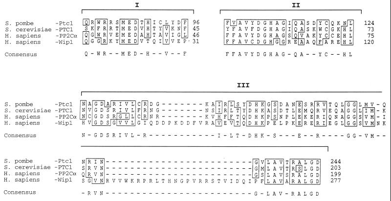
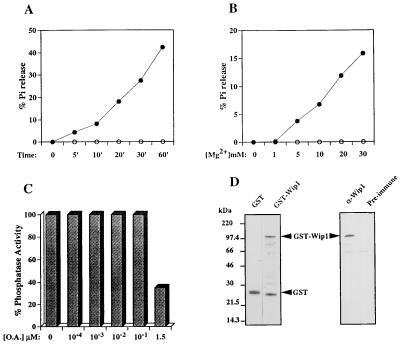
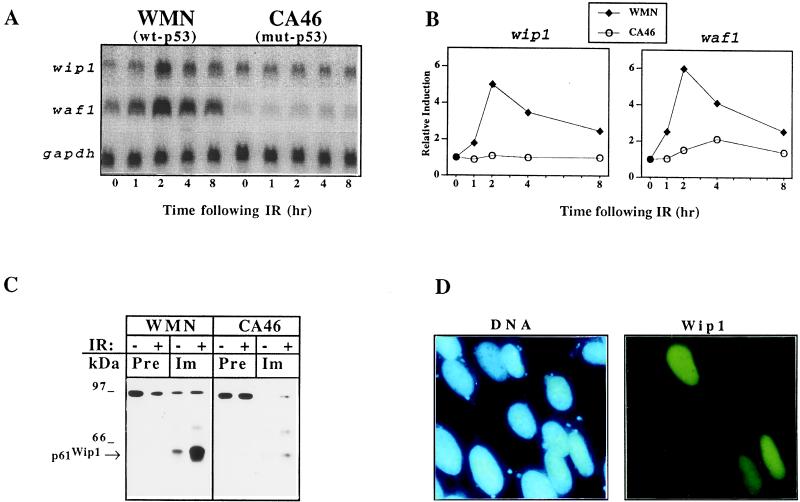
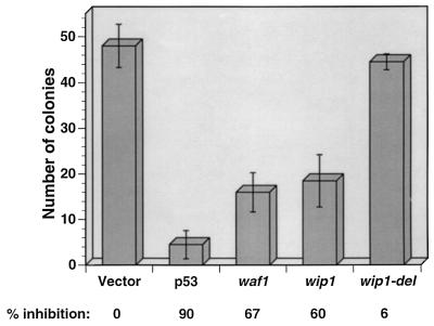
Exposure of mammalian cells to ionizing radiation (IR) induces a complex array of cellular responses including cell cycle arrest and/or apoptosis. IR-induced G1 arrest has been shown to depend on the presence of the tumor suppressor p53, which acts as a transcriptional activator of several genes. p53 also plays a role in the induction of apoptosis in response to DNA damage, and this pathway can be activated by both transcription-dependent and -independent mechanisms. Here we report the identification of a novel transcript whose expression is induced in response to IR in a p53-dependent manner, and that shows homology to the type 2C protein phosphatases. We have named this novel gene, wip1. In vitro, recombinant Wip1 displayed characteristics of a type 2C phosphatase, including Mg2+ dependence and relative insensitivity to okadaic acid. Studies performed in several cell lines revealed that wip1 accumulation following IR correlates with the presence of wild-type p53. The accumulation of wip1 mRNA following IR was rapid and transient, and the protein was localized to the nucleus. Similar to waf1, ectopic expression of wip1 in human cells suppressed colony formation. These results suggest that Wip1 might contribute to growth inhibitory pathways activated in response to DNA damage in a p53-dependent manner.


Figures

Figure 1
Figure 1
Wip1 partial amino acid sequence and protein sequence alignment. wip1 cDNA has an ORF encoding for a protein of 605 amino acids. The three conserved regions among PP2C are outlined (I, II, and III). The figure shows the amino acid sequence alignment within the three identified conserved regions (25) (sequence analysis software from Genetics Computer Group) between human PP2Cα, Wip1, Ptc1 from Schizosaccharomyces pombe, and PTC1 from Saccharomyces cerevisiae. Identical or conserved amino acids are boxed. Numbers on the right identify the amino acid position for each protein. Wip1 carries sequence features typical of this class of phosphatases.
Figure 2
Figure 2
Biochemical characterization of Wip1 as a PP2C. (A) A typical time course experiment showing time-dependent [32P]casein dephosphorylation in the presence of 20 mM Mg2+ by GST–Wip1 (•) but not by the control GST alone (○). PP2Cs are defined by the requirement for Mg2+ for their activity. B shows that GST–Wip1 phosphatase activity is Mg2+ concentration dependent; no activity was detected in absence of Mg2+, but increased linearly with Mg2+ concentrations ranging from 1–30 mM (incubation time 10 min). Another characteristic of the 2C class of phosphatases is their insensitivity to OA as opposed to PP1 and PP2A. (C) A GST–Wip1 phosphatase assay in presence of 20 mM Mg2+ and increasing concentrations of OA. GST–Wip1 is relatively insensitive to OA concentrations that inhibit PP1 and PP2A (10–100 nM). However, at higher concentrations (1.5 μM) GST–Wip1 activity shows partial inhibition by OA. D Left, shows a Coomassie blue-stained polyacrylamide gel of the purified GST–Wip1 and GST used in the phosphatase assays; on the right, a Western blot of the purified GST–Wip1 protein using a polyclonal antisera raised against a Wip1 carboxy-terminal peptide (GQKKIGNPLLHQRKT) (α-GST–Wip1). Preimmune sera was used as a control. Bands lower than full-length GST–Wip1 detected by Coomassie staining (Fig. 3D Left) are degradation products of the full-length protein and can be detected by Western blot analysis using polyclonal antisera raised against the GST–Wip1 fusion protein (data not shown).
Figure 3
Figure 3
Analysis of wip1 expression following IR. (A) wip1 mRNA levels increase after IR in WMN cells and reaches its peak of expression at 2 hr with a time course of induction very similar to waf1, previously shown to be directly induced by wt-p53 (8). No induction was detected in CA46 either for wip1 or for waf1, consistent with the presence of mut-p53 in this cell line. (B) Quantitation of the Northern blot shown in A by using a PhosphorImager model 425 (Molecular Dynamics). (C) Western blot of Wip1 immunoprecipitated from the nuclear fraction of WMN and CA46 cells before and 4 hr after exposure to 6.3 Gy (− or + IR). Detectable amounts of Wip1 were present only in the nuclear fraction of WMN (immunoprecipitation/Western blot of cytosolic fraction not shown), and its levels increased following IR. Immunoprecipitation was performed using either preimmune sera or antisera raised against GST–Wip1. (D) DNA and Wip1 staining of Saos-2 cells transiently transfected with a cDNA encoding for Wip1-FLAG. The fluorescein staining, which is specific for the transfected cells, can be superimposed onto the 4′-6 diamidino-2-phenylindole staining of DNA of the corresponding cells, thus confirming the nuclear localization of Wip1.
Figure 4
Figure 4
Wip1 inhibits cell growth. T98G cells were transfected with either pCMV-neo, or pCMV-neo containing p53, waf1, wip1, and wip1-del cDNAs. Following transfection cells were selected in medium containing G418 (800 μg/ml), and two weeks later colonies were scored. Shown are the mean values ± SD of three different experiments.

References

    1. Hollestein M, Sidransky D, Vogelstein B, Harris C C. Science. 1991;253:49–53. - PubMed
    1. Bates S, Vousden K H. Curr Opin Genet Dev. 1996;6:12–19. - PubMed
    1. Donehower L A, Harvey M, Slagle B L, McArthur M J, Montgomery C A, Jr, Butel J S, Bradley A. Nature (London) 1992;356:215–221. - PubMed
    1. Kastan M B, Zhan Q, El-Deiry W S, Carrier F, Jacks T, Walsh W V, Plunkett B S, Vogelstein B, Fornace A J. Cell. 1992;71:587–597. - PubMed
    1. Lowe S W, Schmitt S W, Smith B A, Osborne B A, Jacks T. Nature (London) 1993;362:847–849. - PubMed

Publication types

MeSH terms

Associated data