. 1997 Jun 10;94(12):6087-92.
doi: 10.1073/pnas.94.12.6087.

Iron-sulfur cluster disassembly in the FNR protein of Escherichia coli by O2: [4Fe-4S] to [2Fe-2S] conversion with loss of biological activity

Affiliations

Iron-sulfur cluster disassembly in the FNR protein of Escherichia coli by O2: [4Fe-4S] to [2Fe-2S] conversion with loss of biological activity

N Khoroshilova et al. Proc Natl Acad Sci U S A. .

Abstract

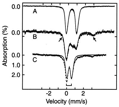
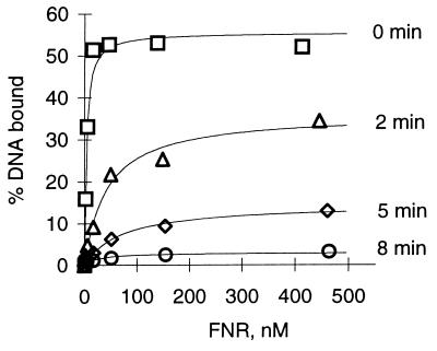
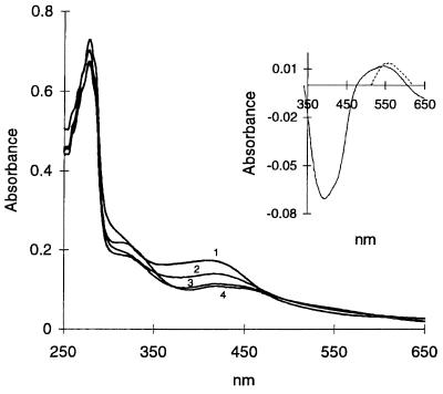
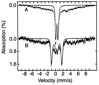
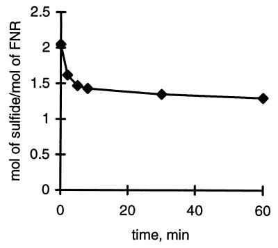
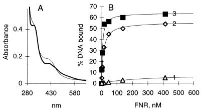
The transcription factor FNR (fumarate nitrate reduction) requires the presence of an iron-sulfur (Fe-S) cluster for its function as a global transcription regulator in Escherichia coli when oxygen becomes scarce. To define the oxidation state and type of Fe-S cluster present in the active form of FNR, we have studied anaerobically purified FNR with Mössbauer spectroscopy. Our data showed that this form of FNR contained a [4Fe-4S]2+ cluster (delta = 0.45 mm/s; DeltaEQ = 1.22 mm/s) and that the [4Fe-4S]2+ cluster was rapidly destroyed on exposure of FNR to air. Under these conditions, the yellow-green active form of FNR turned deep red; analysis of sulfide indicated that 70% of the labile sulfide was still present, suggesting that the Fe-S cluster had been converted into a different form. Little [3Fe-4S] cluster was, however, detected by EPR. According to Mössbauer spectroscopy, the [4Fe-4S]2+ cluster was converted in about 60% yield to a [2Fe-2S]2+ cluster (delta = 0.28 mm/s; DeltaEQ = 0.58 mm/s) following 17 min of exposure to air. The [2Fe-2S]2+ cluster form of FNR was much more stable to oxygen, but was unable to sustain biological activity (e.g., DNA binding). However, DNA binding and the absorption spectrum characteristic of the [4Fe-4S]2+ cluster could be largely restored from the [2Fe-2S]2+ form when Cys, Fe, DTT, and the NifS protein were added. It has yet to be determined whether the form of FNR containing the [2Fe-2S]2+ cluster has any biological significance, e.g., as an in vivo intermediate that is more rapidly converted to the active form than the apoprotein.


Figures

Figure 1
Figure 1
The 4.2 K Mössbauer spectra of anaerobically isolated FNR (A), and FNR exposed to oxygen for 2 min (B) and 17 min (C). The solid line in A is a doublet for [4Fe-4S]2+ simulated with ΔEQ = 1.22 mm/s and δ = 0.45 mm/s. The solid line in C is a simulated spectrum using one doublet with ΔEQ = 0.58 mm/s and δ = 0.28 mm/s for the [2Fe-2S]2+ cluster (bracket). The solid line drawn through the data in B is a superposition of the theoretical curves for the [4Fe-4S]2+ cluster and the [2Fe-2S]2+ cluster, plus a contribution (arrows) from the Fe2+ component. The relative proportions of the various species are listed in Table 1.
Figure 2
Figure 2
Effect of oxygen on DNA binding by FNR. Assays for DNA binding were carried out under anaerobic conditions and quantified by measuring the percentage of the 48-bp DNA fragment bound by FNR as a function of protein concentration. Anaerobic protein (0 min) was exposed to air for 2, 5, and 8 min as described, prior to evacuating and flushing with argon gas.
Figure 3
Figure 3
Absorption spectra of wild-type FNR before and after exposure to air. The spectrum of FNR (1.0 mg/ml) was recorded under anaerobic conditions (1) and after uncapping and mixing the cuvette contents with air for 1 min (2), 10 min (3), and 60 min (4). (Inset) Shows the difference spectrum of oxygen-exposed FNR (60 min) minus anaerobic FNR protein (solid line) and the difference spectrum of a [2Fe-2S] model cluster minus that of a [4Fe-4S] model cluster (broken line) replotted from Fig. 2 of ref. 16 in arbitrary units.
Figure 4
Figure 4
Mössbauer spectra of FNR exposed for 17 min to oxygen, recorded at 4.2 K in zero field (A) and a field of 8.0 T applied parallel to the observed γ radiation. The solid line in A is a doublet outlining the contribution of the [2Fe-2S]2+ cluster. The solid line in B is a theoretical spectrum computed with values of ΔEQ and δ obtained in zero field with the assumption that both (equivalent) Fe sites of the cluster reside in a diamagnetic environment. Theoretical curves are drawn to emphasize hyperfine splittings; they do not represent correct spectral areas.
Figure 5
Figure 5
Loss of sulfide from FNR upon exposure to air. Disappearance of inorganic sulfide as a function of time of exposure of FNR to air. The reaction with air was quenched as described in Materials and Methods.
Figure 6
Figure 6
Reconstitution of the [4Fe-4S] cluster in FNR from the [2Fe-2S] cluster. (A) Absorption spectrum of air-oxidized FNR (thick line) and the same after reconstitution of the [4Fe-4S] cluster with Azotobacter vinelandii NifS protein as described (thin line). The reference cuvette contained the same mixture except for FNR. (B) DNA binding of air-oxidized (10 min) FNR lacking the 4Fe cluster (1), air-oxidized FNR following reconstitution of the [4Fe-4S] cluster (2), and FNR as obtained on purification (3).

References

    1. Spiro S. Antonie van Leeuwenhoek. 1994;66:23–36. - PubMed
    1. Khoroshilova N, Beinert H, Kiley P J. Proc Natl Acad Sci USA. 1995;92:2499–2503. - PMC - PubMed
    1. Lazazzera B A, Beinert H, Khoroshilova N, Kennedy M C, Kiley P J. J Biol Chem. 1996;271:2762–2768. - PubMed
    1. Green J, Bennett B, Jordan P, Ralph E T, Thomson A J, Guest J R. Biochem J. 1996;316:887–892. - PMC - PubMed
    1. Green J, Trageser M, Six S, Unden G, Guest J R. Proc R Soc London B. 1991;244:137–144. - PubMed

Publication types

MeSH terms

LinkOut - more resources