

Three-dimensional diffuse x-ray scattering from crystals of Staphylococcal nuclease
- PMID: 9177191
- PMCID: PMC21023
- DOI: 10.1073/pnas.94.12.6180
Three-dimensional diffuse x-ray scattering from crystals of Staphylococcal nuclease
Abstract
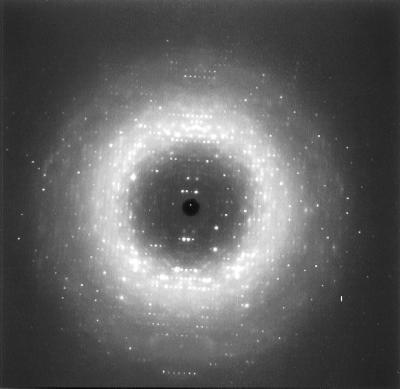
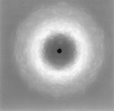
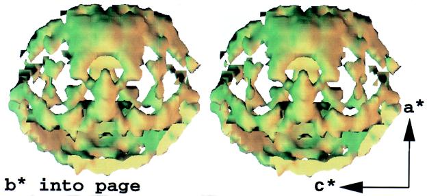
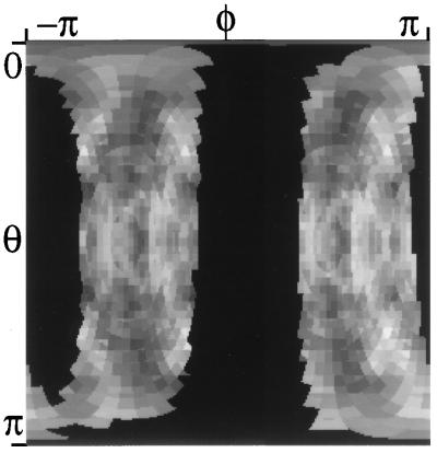
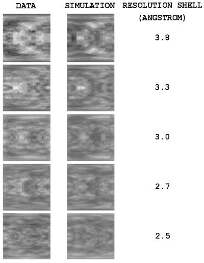
We have developed methods for obtaining and characterizing three-dimensional maps of the reciprocal-space distribution of diffuse x-ray scattering from protein crystals, and have used the methods to study the nature of disorder in crystals of Staphylococcal nuclease. Experimentally obtained maps are 99.5% complete in the reciprocal-space resolution range of 10 A-2.5 A, show symmetry consistent with the P41 space group of the unit cell, and are highly reproducible. Quantitative comparisons of the data with three-dimensional simulations imply liquid-like motions of the protein [Caspar, D. L. D., Clarage, J., Salunke, D. M. & Clarage, M. (1988) Nature (London) 332, 659-662], with a correlation length of 10 A and a root-mean-square displacement of 0.36 A.
Figures
References
-
- Debye P. Verh Dtsch Phys Ges. 1913;15:678. , 738, 857.
-
- Faxén H. Z Phys. 1923;17:266–278.
-
- Waller I. Ph.D. thesis. Uppsala, Sweden: Uppsala University; 1925.
-
- Laval J. Bull Soc Franc Min. 1939;62:137–253.
-
- Born M, Sarginson K. Proc R Soc London Ser A. 1941;179:69–93.
Publication types
MeSH terms
Substances
LinkOut - more resources
Full Text Sources
