. 1997 Jun 10;94(12):6191-6.
doi: 10.1073/pnas.94.12.6191.

Interaction of protein kinase C zeta with ZIP, a novel protein kinase C-binding protein

Affiliations

Interaction of protein kinase C zeta with ZIP, a novel protein kinase C-binding protein

A Puls et al. Proc Natl Acad Sci U S A. .

Abstract

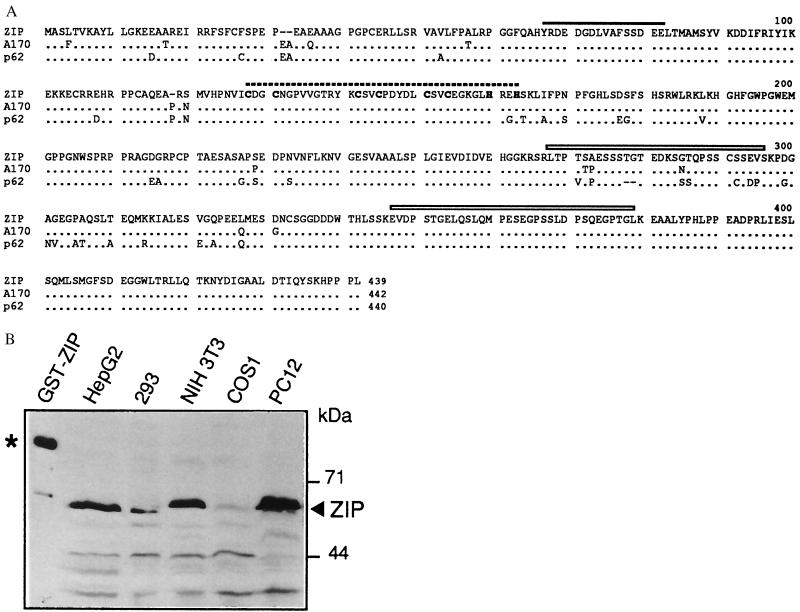
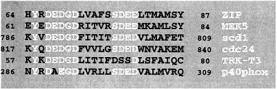
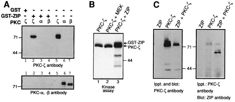
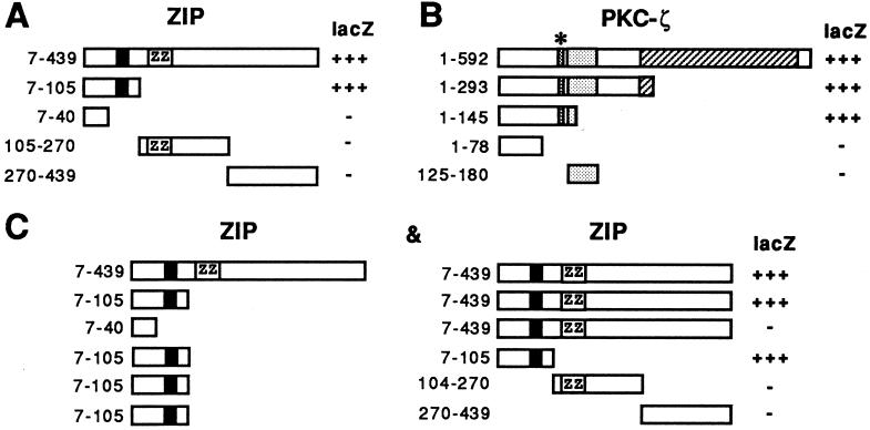
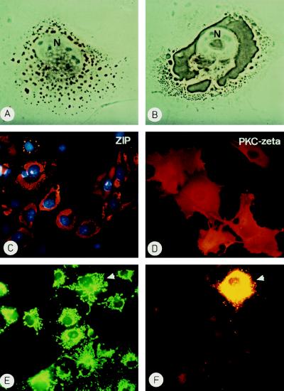
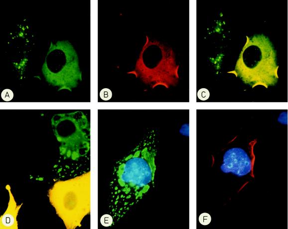
The atypical protein kinase C (PKC) member PKC-zeta has been implicated in several signal transduction pathways regulating differentiation, proliferation or apoptosis of mammalian cells. We report here the identification of a cytoplasmic and membrane-associated protein that we name zeta-interacting protein (ZIP) and that interacts with the regulatory domain of PKC-zeta but not classic PKCs. The structural motifs in ZIP include a recently defined ZZ zinc finger as a potential protein binding module, two PEST sequences and a novel putative protein binding motif with the consensus sequence YXDEDX5SDEE/D. ZIP binds to the pseudosubstrate region in the regulatory domain of PKC-zeta and is phosphorylated by PKC-zeta in vitro. ZIP dimerizes via the same region that promotes binding to PKC-zeta suggesting a competitive situation between ZIP:ZIP and ZIP:PKC-zeta complexes. In the absence of PKC-zeta proper subcellular localization of ZIP is impaired and we show that intracellular targeting of ZIP is dependent on a balanced interaction with PKC-zeta. Taking into account the recent isolation of ZIP by others in different contexts we propose that ZIP may function as a scaffold protein linking PKC-zeta to protein tyrosine kinases and cytokine receptors.


Figures

Figure 1
Figure 1
Cloning of a ZIP cDNA. (A) Alignment of ZIP with murine sequence A170 (30) and human protein p62 (19). The Cdc24 homology (see Fig. 2) is marked by a solid line, the ZZ finger by a broken line and the PEST sequences by boxes. Dots represent identities, dashes mark gaps in the alignment. (B) Expression of ZIP was demonstrated by Western blot analysis in five cell lines as indicated. As a control for immunostaining a GST-ZIP fusion protein (93 kDa) is shown in lane 1 and marked with an asterisk.
Figure 2
Figure 2
A putative protein binding module in ZIP. Homologies were identified with the blast program (32) and sequences were extracted from the following references: S. pombe scd1 (33), S. cerevisiae Cdc24 (34), rat MEK5 (35), human TRK-T3 (36), and human p40phox (37).
Figure 3
Figure 3
Specific interaction between ZIP and PKC-ζ. (A) GST control protein or a GST-ZIP fusion protein (as indicated) was immobilized on glutathione-Sepharose beads and incubated with purified PKC-ζ (lane 2), with purified PKC-α (lane 3) or purified PKC-βI (lane 4). After washing the complexes were resolved by SDS/PAGE and the same immunoblot was sequentially incubated with antiserum against PKC-ζ (top) or PKC-α and -β (bottom). Lanes 5–7 show the amounts of purified PKC proteins used in the binding assays. (B) PKC-ζ purified from insect cells was incubated in a phosphorylation reaction with [32P]-labeled ATP either alone (lane 1), with purified GST-ZIP (lane 3) or with an unrelated control protein GST-MEK (lane 2). Shown is an autoradiograph of the phosphorylation reaction. (C) PKC-ζ was immunoprecipitated from extracts of COS cells expressing ZIP (lane 1), PKC-ζ (lane 2), or both ZIP and PKC-ζ (lane 3). The immunoprecipitates were analyzed for the presence of PKC-ζ (Left) or ZIP (Right) by sequential Western blot analysis.
Figure 4
Figure 4
Binding sites on ZIP and PKC-ζ. (A) Deletion mutants of ZIP as shown were coexpressed in HF7c yeast cells together with full-length PKC-ζ. Staining in the β-galactosidase assay is indicated on the right (lacZ). Numbers on the left give amino acid positions. (B) Deletion mutants of PKC-ζ as shown were analyzed together with full-length ZIP as in A. (C) ZIP mutants fused to the GAL4 activation domain (Left) were coexpressed in HF7c yeast cells with mutants of ZIP fused to the GAL4 DNA-binding domain (Right). β-Galactosidase activity of the interaction of the respective pair (Left and Right) is indicated on the right (lacZ). The black box in ZIP represents the Cdc24 homology (see Fig. 2), the relative position of the ZZ zinc finger is indicated by ZZ. The stippled box in PKC-ζ denotes the PKC-ζ zinc finger that is preceded by the pseudosubstrate sequence marked by an asterisk.
Figure 5
Figure 5
Ectopic expression of ZIP or PKC-ζ. COS cells transfected with an expression vector pMT2-ZIP were fixed after 24 hr (A) or 48 hr (B and C). ZIP was detected by indirect immmunochemistry using a horseradish peroxidase-coupled (A and B) or Texas Red-labeled (C) secondary antibody. N marks the nucleus in A and B and nuclei in C were stained with 4′,6-diamidino-2′-phenylindole. (D) COS cells transfected with a pMT2-PKC-ζ expression vector were stained for PKC-ζ at 48 hr posttransfection with a Texas Red-labeled secondary antibody. (E and F) COS cells expressing ZIP were incubated with rhodamine-123 at 40 hr to stain mitochondria (E), then they were fixed and ZIP expression was detected by indirect immunofluorescence (F). The cell expressing ZIP is marked by an arrowhead.
Figure 6
Figure 6
Intracellular targeting of ZIP by PKC-ζ but not by ERK. COS cells were cotransfected with expression vectors encoding myc-tagged ZIP and PKC-ζ (A–D) or myc-ZIP and ERK2 (E and F). After 40 hr cells were fixed and proteins were visualized by indirect immunofluorescence. ZIP was detected by fluoresceine (green), PKC-ζ, and ERK by rhodamine (red). Double exposures (C and D) (yellow) show the overlapping fluorescence of ZIP and PKC-ζ. (A–C) The left cell only expresses ZIP and shows punctate staining, the right cell coexpresses ZIP and PKC-ζ and shows diffuse staining of ZIP. (D) In the two yellow cells ZIP and PKC-ζ colocalize, the cell at the top right only expresses ZIP. (E and F) Despite coexpression of the unrelated protein kinase ERK (red) ZIP (green) does not show even distribution but is still aggregated. Nuclei are stained with 4′,6-diamidino-2′-phenylindole.

References

    1. Nishizuka Y. Science. 1992;258:607–614. - PubMed
    1. Hug H, Sarre T F. Biochem J. 1993;291:329–343. - PMC - PubMed
    1. Stabel S. Semin Cancer Biol. 1994;5:277–284. - PubMed
    1. Kiley S C, Jaken S, Whelan R, Parker P J. Biochem Soc Trans. 1995;23:601–605. - PubMed
    1. Nakamura S, Nishizuka Y. J Biochem (Tokyo) 1994;115:1029–1034. - PubMed

Publication types

MeSH terms

Associated data