. 1997 Jun 10;94(12):6210-5.
doi: 10.1073/pnas.94.12.6210.

Aberrant retention of tyrosinase in the endoplasmic reticulum mediates accelerated degradation of the enzyme and contributes to the dedifferentiated phenotype of amelanotic melanoma cells

Affiliations

Aberrant retention of tyrosinase in the endoplasmic reticulum mediates accelerated degradation of the enzyme and contributes to the dedifferentiated phenotype of amelanotic melanoma cells

R Halaban et al. Proc Natl Acad Sci U S A. .

Abstract

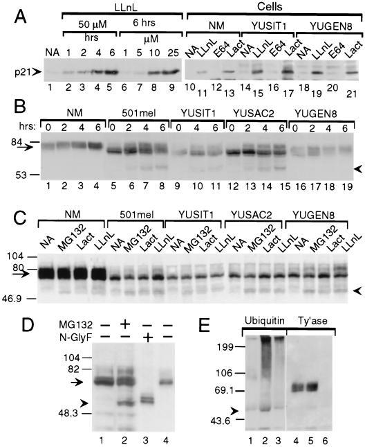
The loss of tyrosinase, the key enzyme in melanin synthesis, has been implicated in the dedifferentiation of malignant melanocytes. The presence of tyrosinase transcripts and antigenic peptides in melanoma tumors prompted us to investigate whether the basis for the loss of the enzyme was proteolytic degradation. Toward this aim, we followed the kinetics of synthesis, degradation, processing, chaperone binding, inhibitor sensitivity, and subcellular localization of tyrosinase in normal and malignant melanocytes. We found that, in amelanotic melanoma cell lines, tyrosinase failed to reach the melanosome, the organelle for melanin synthesis, because it was retained in the endoplasmic reticulum (ER) and then degraded. Tyrosinase appeared mostly as a 70-kDa core-glycosylated, endoglycosidase H-sensitive, immature form bound to the ER chaperone calnexin and had a life-span of only 25% of normal. Maturation and transit from the ER to the Golgi compartment was facilitated by lowering the temperature of incubation to 31 degrees C. Several proteasome inhibitors caused the accumulation of an approximately 60-kDa tyrosinase doublet that was more prominent in malignant than in normal melanocytes and promoted, to various degrees, the maturation of tyrosinase in melanoma cells and the translocation of the enzyme to melanosomes. The appearance of ubiquitinated tyrosinase after treatment of normal melanocytes with N-acetyl-L-leucinyl-L-leucinal-L-norleucinal reinforced our notion that some tyrosinase is normally degraded by proteasomes. Proteolysis of tyrosinase by proteasomes is consistent with the production of antigenic tyrosinase peptides that are presented to the immune system by major histocompatibility complex class I molecules.


Figures

Figure 1
Figure 1
Tyrosinase processing and degradation in normal and malignant melanocytes. (A) Steady-state tyrosinase protein. Anti-tyrosinase (polyclonal) Western blots of glycoproteins from normal melanocytes of individual donors (lanes 1–10), mixed melanocytes from six neonatal donors (mix, lane 11), or metastatic amelanotic melanoma cell lines 501 mel, YUSIT1, YUSAC2, and the slightly melanotic YUGEN8 (lanes 12–15). The normal adult melanocytes were from two healthy volunteers (lanes 7 and 8), two patients with vitiligo (lanes 6 and 9), and one patient with mastocytosis (lane 10). Two of the donors had a light complexion, blond hair, and blue eyes and were unable to tan well (lanes 7 and 9). (B) Kinetics of tyrosinase processing and degradation. Autoradiograms of radiolabeled proteins immunoprecipitated with anti-tyrosinase antibodies (lanes 1–14) or control rabbit IgG (lane 15). Cultures pulsed for 15 minutes with 35S were harvested immediately (lanes marked 0) or after a chase with regular medium for the duration of 0.25, 0.5, 1, 3, 6, or 10 h. Immunoprecipitation reaction mixtures contained 3.6 × 10⁷ cpm in trichloroacetic acid-precipitable material (lanes 1–11) or 2 × 10⁷ cpm (lanes 12–15). X-ray films were exposed to the dried gels for 4 days. (C) Band densities as a function of chase time. Data were derived from the x-ray films presented in B, scanned with a Molecular Dynamics Image Analyzer. ○, Lanes 5–8; ▴, lanes 9–11; and ▪, lanes 12–14, normalized to compensate for lower specific radioactivity in labeled proteins.
Figure 4
Figure 4
Evidence for tyrosinase degradation by the proteasome–ubiquitin pathway. Western blots with anti-p21 (A), anti-tyrosinase (polyclonal, B; monoclonal, C–E) or anti-ubiquitin (E) antibodies. (A) Proteasome inhibitors block p21WAF1/Cip1 degradation. Accumulation of p21 in response to LLnL in metastatic melanoma line YUPAC7 as a function of time and concentration (lanes 1–9) and in normal melanocytes (NM) and melanoma cell lines after treatment for 4 h with LLnL (50 μM) or with lactacystin (Lact, 60 μM) but not with DMSO (0.1%, NA) or E64 (50 μM) (lanes 10–21). (B) Accumulation of 60-kDa tyrosinase doublet and increased efficiency of tyrosinase processing in response to LLnL. Shown is tyrosinase in whole cell lysates (40 μg/lane) from normal melanocytes (NM) or melanoma cells treated with DMSO for 6 h (lanes marked 0) or LLnL (50 μM) for 2, 4, or 6 h. (C) Accumulation of 60-kDa tyrosinase doublet by additional inhibitors. Cells were treated for 3 h with DMSO (NA), MG132 (50 μM), lactacystin (Lact, 30 μM), or LLnL (50 μM). (D) The 60-kDa band comigrates with deglycosylated tyrosinase. Lanes: 1 and 2, tyrosinase in whole cell lysates from 501mel cells treated for 6 h with DMSO (−) or MG132 (+, 50 μM); 3 and 4, lactin-bound glycoproteins from 501mel cells after overnight incubation without (−) or with N-glycosidase F (N-glyF, +). (E) Ubiquitination of tyrosinase. Products of immunoprecipitations with anti-tyrosinase polyclonal antibodies (lanes 1, 2, 4, and 5) or control rabbit IgG (lanes 3 and 6) were Western blotted with anti-ubiquitin 4F3 mAb. After stripping, the same membrane was probed with anti-tyrosinase mAb (Ty’ase). Extracts were from normal melanocytes incubated for 4 h with DMSO (lanes 1 and 4) or LLnL (50 μM, lanes 2, 3, 5, and 6). Arrowheads in B–E point at the 60-kDa doublet of tyrosinase. Arrows point at normal and melanoma tyrosinase glycoproteins.
Figure 3
Figure 3
Recovery of mature tyrosinase after culturing melanoma cells at reduced temperature. (A and B) Anti-tyrosinase (mAb) Western analysis of glycoproteins derived from cells grown at 37°C or exposed to 31°C for increasing durations (A) or 24 h (B), not subjected to (−) or subjected to (+) Endo H digestion. (Note: The tyrosinase glycoforms of 501 mel cells at 37°C are presented in Fig. 2A.) (C) Autoradiogram of metabolically labeled radioactive tyrosinase derived from cells pulsed for 15 min and harvested immediately (lanes 1 and 6) or after a chase of 1 or 3 h (lanes 2–5 and 7–10 as indicated). Immunoprecipitations with anti-tyrosinase antibodies (lanes 1–8) or control antibodies (lanes 9 and 10); gels were exposed on x-ray film for 5 days. (D) Tyrosinase activity in normal and malignant melanocytes grown at 37°C (37) or after exposure for 4 h to 31°C (31). The SE in tyrosinase activity of duplicate samples ranged from 3 to 10% of total. (Inset) Results with melanoma cell lines are shown on a smaller scale to highlight the different enzyme activity.
Figure 5
Figure 5
Further evidence that LLnL enhances maturation of tyrosinase, leading to translocation to prememalnosomes. (A) Autoradiogram of radioactive proteins immunoprecipitated with tyrosinase antibodies (lanes 1–21) or control rabbit IgG (lane 22). Cells were pulsed with 35S for 15 min and harvested immediately (0) or after incubation for the indicated periods of time in regular medium. LLnL was either absent (−) or present (+) during the incubation–chase period. (B) LLnL promotes tyrosinase–calreticulin complexing. Western blot of calreticulin-immune precipitates probed with anti-tyrosinase mAb. Lysates for immunoprecipitation were prepared from cells treated with DMSO (−) or LLnL (50 μM for 4 h) (+). (C) LLnL enhances translocation of tyrosinase into premelanosomes. Electron micrographs depict dihydroxy l-phenyl alanine-(tyrosinase) reactivity in untreated (a, d) or LLnL-treated (50 μM for 4.5 h; b, c, and e) amelanotic YUSAC2 melanoma cells. Solid arrowheads indicate electron-opaque reaction product. Insets d and e show unreactive (open arrow) and reactive (solid arrowhead) premelanosomes. Dopa-reactive, trans-Golgi elements (small arrowheads) in an LLnL-treated cell in c. n = nucleus. (Bars = 1 μm in a, b, and c and 0.5 μm in d and e.)
Figure 2
Figure 2
Arrest of tyrosinase in the ER of amelanotic melanoma cells. (A) Endo H-resistant forms were not detected in melanoma cells. The anti-tyrosinase (polyclonal) Western blot shows nondigested (−) or Endo H-digested (+) tyrosinase from normal (NM) and malignant melanocytes. (B) Reducing agents induced ER retention in normal but not malignant melanocytes. Anti-tyrosinase (mAb) Western analysis of undigested (−) or Endo H-digested (+) glycoproteins derived from cells treated for 4 h with 2-mercaptoethanol (2-ME) or DTT, 5 mM each. (C) Tyrosinase of melanoma cells associated with the ER chaperone calnexin but not calreticulin. Cell extracts from normal (lanes 1 and 6) or malignant melanocytes (lanes 2–5 and 7–8) were subjected to immunoprecipitation with polyclonal antibodies against calnexin (lanes 1–5) or calreticulin (lanes 6–10), followed by Western blotting with an mAb against tyrosinase (representative experiment of two).

References

    1. Halaban R, Pomerantz S H, Marshall S, Lambert D T, Lerner A B. J Cell Biol. 1983;97:480–488. - PMC - PubMed
    1. Müller G, Ruppert S, Schmid E, Schütz G. EMBO J. 1988;7:2723–2730. - PMC - PubMed
    1. Halaban R. Semin Cancer Biol. 1993;4:171–181. - PubMed
    1. Eberle J, Garbe C, Wang N P, Orfanos C E. Pigm Cell Res. 1995;8:307–313. - PubMed
    1. Mellado B, Colomer D, Castel T, Muiloz M, Carballo E, Galon M, Mascaro J M, Vives-Corrons J L, Grou J J, Estape J. J Clin Oncol. 1996;14:2091–2097. - PubMed

Publication types

MeSH terms