. 1997 Jun 10;94(12):6222-7.
doi: 10.1073/pnas.94.12.6222.

Intracellular structures of normal and aberrant Plasmodium falciparum malaria parasites imaged by soft x-ray microscopy

Affiliations

Intracellular structures of normal and aberrant Plasmodium falciparum malaria parasites imaged by soft x-ray microscopy

C Magowan et al. Proc Natl Acad Sci U S A. .

Abstract

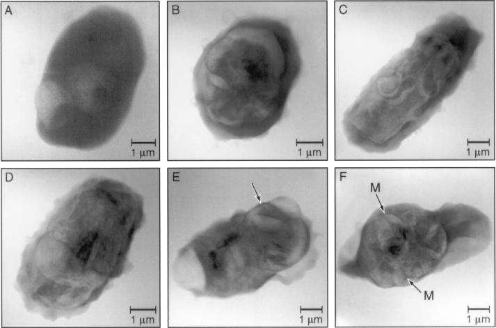
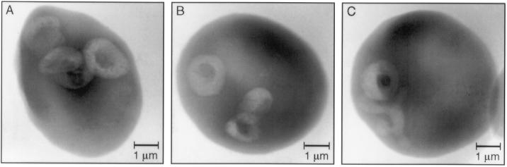
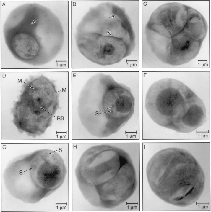
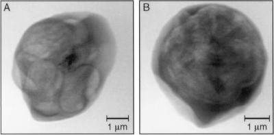
Soft x-ray microscopy is a novel approach for investigation of intracellular organisms and subcellular structures with high spatial resolution. We used x-ray microscopy to investigate structural development of Plasmodium falciparum malaria parasites in normal and genetically abnormal erythrocytes and in infected erythrocytes treated with cysteine protease inhibitors. Investigations in normal red blood cells enabled us to recognize anomalies in parasite structures resulting from growth under unfavorable conditions. X-ray microscopy facilitated detection of newly elaborated structures in the cytosol of fixed, unstained, intact erythrocytes, redistribution of mass (carbon) in infected erythrocytes, and aberrant parasite morphology. In cysteine protease inhibitor-treated, infected erythrocytes, high concentrations of material were detected in abnormal digestive vacuoles and aggregated at the parasite plasma membrane. We have demonstrated that an abnormal host erythrocyte skeleton affects structural development of parasites and that this aberrant development can be detected in the following generation when parasites from protein 4.1-deficient red blood cells infect normal erythrocytes. This work extends our current understanding of the relationship between the host erythrocyte membrane and the intraerythrocytic malaria parasite by demonstrating for the first time that constituents of the erythrocyte membrane play a role in normal parasite structural development.


Figures

Figure 3
Figure 3
Soft x-ray micrographs of P. falciparum malaria parasites infecting protein 4.1-deficient erythrocytes. (A) Two ring stage parasites infecting an elliptocytic erythrocyte. Structurally there are not obvious differences at the ring stage between parasites in normal and protein 4.1-deficient erythrocytes. Note the parasite apposed to erythrocyte membrane protuberance (exposure, 30 sec). (B–E) Trophozoites show structural derangements during maturation in protein 4.1-deficient erythrocytes. Parasites have a prolate spheroidal form (C and D) and complicated structures with redistributed mass not detected in trophozoites infecting normal erythrocytes. There is obvious damage to the erythrocyte membranes including (E), an image of a parasite that does not express major malarial proteins that associate with the erythrocyte membrane. Note the sharp edge (arrow) in E, demonstrating the resolving power of our microscope. The width of this edge is <80 nm (exposure: B, C, and E, 20 sec; D, 60 sec). (F) Multinucleated schizont in a protein 4.1-deficient erythrocyte. Arrangement of forming merozoites (M) and the residual body appears more disorganized than in normal erythrocytes (exposure, 30 sec).
Figure 1
Figure 1
Soft x-ray micrographs of intraerythrocytic ring stage P. falciparum malaria parasites imaged in normal erythrocytes. (A and B) Multiply infected erythrocytes from synchronized in vitro cultures were collected at the initiation of the experiment, washed three times in filtered PBS, and fixed in 1% or 2% glutaraldehyde. Ring stage parasites appeared discoidal and stippled. The darker area in the center of each parasite indicates more mass in the digestive vacuole (30 sec exposures). (C) An image of a singly infected erythrocyte removed from culture and fixed 6 hr after A and B shows a region of lower mass, compared with the erythrocyte cytosol, which extends from the parasite into the erythrocyte cytosol. This newly elaborated structure has not been seen before in fixed, unlabeled intact cells (exposure, 60 sec).
Figure 2
Figure 2
Soft x-ray micrographs of intraerythrocytic trophozoite stage P. falciparum malaria parasites imaged in normal erythrocytes: untreated and treated with cysteine protease inhibitors. (A) An untreated trophozoite 12 hr after initiation of experiment. Note increase in size and structural complexity, and ellipsoidal form. Mass in the erythrocyte cytosol has been redistributed from brighter area (less mass) to dense region (indicated by arrow) surrounding the parasite (exposure, 30 sec). (B) Trophozoite at 30 hr has obvious digestive vacuole containing hemozoin (malarial pigment). Redistribution of hemoglobin at this time point is indicated by the brighter erythrocyte cytosol (less mass) and the dark, highly absorptive material in digestive vacuole. Arrows indicate dense regions surrounding the parasite (exposure, 60 sec). (C) Multiply infected erythrocyte at 36 hr (exposure, 30 sec). (D) Multinucleated schizont at 36 hr. Individual merozoites (M) can be distinguished and the residue of the digestive vacuole [the residual body (RB)] is centrally located. Note advanced state of disintegration of erythrocyte membrane (exposure, 30 sec). (E and F) Leupeptin and (G) ZFR-treated trophozoites exhibit enlarged digestive vacuoles with extremely dense contents. Dense spheres (S) that appear to be in the parasite cytoplasm can be seen. The region surrounding the trophozoite is sheet-like and very dense, while the erythrocyte cytosol is depleted of hemoglobin (exposure, 30 sec). (H and I) ZFR-treated trophozoites exhibit unusual structures and disordered distribution of mass compared with untreated trophozoites. Cleft-like forms and apparently partitioned areas within parasites can be seen (exposure, 30 sec).
Figure 4
Figure 4
Soft x-ray micrographs of parasites from protein 4.1-deficient erythrocytes infecting normal erythrocytes. (A and B) Parasites that matured in protein 4.1-deficient erythrocytes in the previous generation retain structural abnormalities first recognized in the abnormal host erythrocytes when subsequently infecting normal erythrocytes. Unusual structures and disordered density are detected (exposure, 50 sec).

References

    1. Meyer-Ilse W, Medecki H, Jochum L, Anderson E, Attwood D, Magowan C, Balhorn R, Moronne M, Rudolph D, Schmahl G. Synchrotron Radiat News. 1995;8:29–33.
    1. Kirz J, Jacobson C, Howells M. Q Rev Biophys. 1995;28:33–130. - PubMed
    1. Schmahl G, Rudolph D, Niemann B, Guttmann P, Thieme J, Schneider G, David C, Diehl M, Wilhein T. Optik. 1993;93:95–102.
    1. Hoffman S L. N Engl J Med. 1996;335:124–126. - PubMed
    1. Luse S A, Miller L H. Am J Trop Med Hyg. 1971;20:655–660. - PubMed

Publication types

MeSH terms

Substances