. 1997 Jun 10;94(12):6307-12.
doi: 10.1073/pnas.94.12.6307.

The peripheral blood fibrocyte is a potent antigen-presenting cell capable of priming naive T cells in situ

Affiliations
Free PMC article

The peripheral blood fibrocyte is a potent antigen-presenting cell capable of priming naive T cells in situ

J Chesney et al. Proc Natl Acad Sci U S A. .
Free PMC article

Abstract

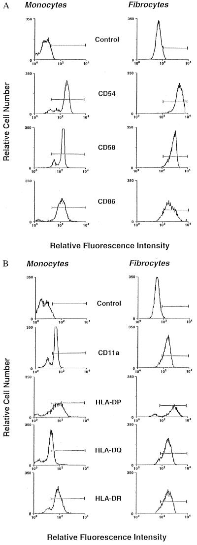
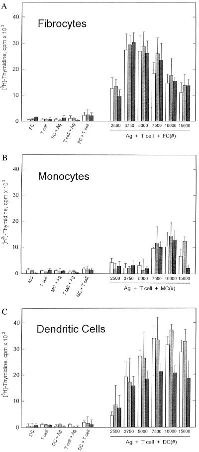
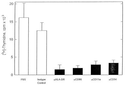
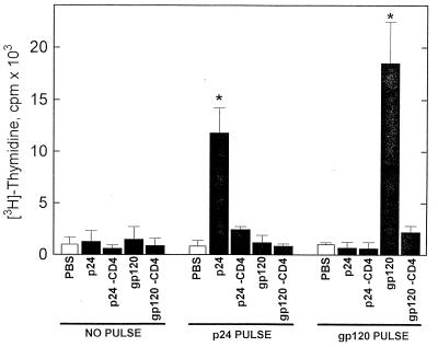
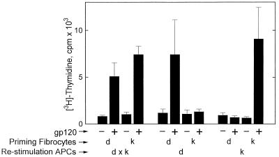
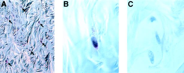
Recent studies have identified a novel population of blood-borne cells, termed fibrocytes, that have a distinct cell surface phenotype (collagen+/CD13(+)/CD34(+)/CD45(+)), rapidly enter sites of tissue injury, and synthesize connective tissue matrix molecules. We found by flow cytometry that purified human fibrocytes express each of the known surface components that are required for antigen presentation, including class II major histocompatability complex molecules (HLA-DP, -DQ, and -DR), the costimulatory molecules CD80 and CD86, and the adhesion molecules CD11a, CD54, and CD58. Human fibrocytes induced antigen-presenting cell-dependent T cell proliferation when cultured with specific antigen and this proliferative activity was significantly higher than that induced by monocytes and nearly as high as that induced by purified dendritic cells. Mouse fibrocytes also were found to express the surface components required for antigen presentation and to function as potent APCs in vitro. Mouse fibrocytes pulsed in vitro with the HIV-proteins p24 or gp120 and delivered to a site of cutaneous injury were found to migrate to proximal lymph nodes and to specifically prime naive T cells. These data suggest that fibrocytes play an early and important role in the initiation of antigen-specific immunity.


Figures

Figure 1
Figure 1
Accessory molecule expression by human fibrocytes and monocytes. Purified, human fibrocytes or monocytes were incubated with phycoerythrin-conjugated (A) or fluorescein isothiocyanate-conjugated (B) mAbs and analyzed by flow cytometry as described. The horizontal line in each panel marks fluorescence intensity greater than the background staining that was observed with an isotype control mAb.
Figure 2
Figure 2
Functional comparison of human fibrocyte, monocyte, and dendritic cell antigen presentation in vitro. Human T cells (2 × 10⁵) purified from a tetanus toxoid-immunized individual were incubated with 2 μg/ml tetanus toxoid together with various numbers of autologous fibrocytes (A), autologous monocytes (B), or autologous dendritic cells (C). After incubation for 4 (□), 5 (▩), and 6 (▪) days, the cultures were pulsed for 12 hr with 4 μCi/ml [3H]thymidine and cell proliferation analyzed by liquid scintillation counting. Controls are illustrated on the left side of each figure: APCs alone (FC, fibrocytes; MC, monocytes; DC, dendritic cells), T cells alone, APCs + Ag (tetanus toxoid), T cells + Ag (tetanus toxoid), and APCs + T cells. Data are expressed as mean ± SD and are representative of one experiment that was performed three times.
Figure 3
Figure 3
Effect of neutralizing anti-HLA-DR, anti-CD86, anti-CD54, or anti-CD11a mAbs on fibrocyte antigen presentation in vitro. Receptor-specific or isotype control (5 μg/ml) mAb were added to a 20:1 autologous T cell:fibrocyte coincubation prior to the addition of tetanus toxoid. The antigen-dependent T cell proliferation assay was conducted as in Fig. 2. Data are expressed as mean ± SD and are representative of one experiment that was performed three times.
Figure 4
Figure 4
Murine fibrocyte antigen presentation in vitro. Murine T cells (2 × 10⁵) purified from the spleens of p24 (A) or gp120 (B) immunized BALB/c were incubated with 2 μg/ml p24 or gp120 together with various numbers of mitomycin C-treated autologous fibrocytes. After incubation for 5 days, the cultures were pulsed for 12 hr with 4 μCi/ml [3H]thymidine and cell proliferation analyzed by liquid scintillation counting. Controls are illustrated on the left side of each figure. Data are expressed as mean ± SD and are representative of one experiment that was performed three times.
Figure 5
Figure 5
Priming of naive T cells in situ with antigen-pulsed murine fibrocytes. Purified BALB/c mouse fibrocytes were cultured for 3 days without (NO PULSE), or with 50 μg/ml of p24 (p24 PULSE), or gp120 (gp120 PULSE) in DME/20% FCS, washed, and injected i.d. (5 × 10⁴ cells) into the right rear footpad. The proximal popliteal lymph nodes were explanted 5 days later, dissociated, and cultured with PBS or 50 μg/ml of p24 or gp120 for 72 hr. To verify the proliferating cell type, CD4+ T cells were depleted by immunomagnetic selection just prior to liquid scintillation counting (p24-CD4, gp120-CD4). The proliferative activity was measured over the last 12 hr of culture by [3H]thymidine incorporation. Data are expressed as mean ± SD and are representative of one experiment that was performed three times.
Figure 6
Figure 6
Requirement of shared MHC haplotype between priming fibrocyte and restimulation APC. DBA-2 × C3H/HeJ F1 mice (H-2dxk) were injected with antigen-pulsed fibrocytes from either parent strain DBA-2, H-2d (d) or C3H/HeJ, H-2k (k). Five days later, the proximal popliteal lymph node cells were isolated and depleted of endogenous class II MHC+ APCs by immunomagnetic selection. APC-depleted lymph node cells (1 × 10⁵) then were cocultured with 1 × 10⁵ mitomycin C-treated F1 (dxk) or parental spleen cells (d or k) as restimulation APCs, with or without gp120. The proliferative activity was measured over the last 12 hr of culture by [3H]thymidine incorporation. Data are expressed as mean ± SD and are representative of one experiment that was performed three times.
Figure 7
Figure 7
Coexpression of CD34 and HLA-DR by cells in a human cutaneous tissue scar. Skin specimens were fixed, sectioned, and labeled with both anti-CD34 mAb and anti-HLA-DR mAb. Binding was detected with an immunoperoxidase-linked (anti-CD34) and an alkaline phosphatase-linked (anti-HLA-DR) secondary antibody. The anti-CD34 positive areas appear brown and granular and the anti-HLA-DR positive regions stain red. The isotype control displays no immunoreactivity. [×200 (A); ×1,000 (B and C).]

References

    1. Mast B A. In: Wound Healing: Biochemical and Clinical Aspects. Cohen I K, Diegelmann R F, Lindblad W J, editors. Philadelphia: Saunders; 1992. pp. 344–355.
    1. Davidson J M. In: Inflammation: Basic Principles and Clinical Correlates. Gallin J I, Goldstein I M, Snyderman R, editors. New York: Raven; 1992. pp. 809–819.
    1. Chu T, Jaffe M B. Br J Cancer Suppl. 1994;23:S4–S10. - PMC - PubMed
    1. Bucala R, Spiegel L A, Chesney J A, Hogan M, Cerami A. Mol Med. 1994;1:71–81. - PMC - PubMed
    1. Freudenthal P S, Steinman R M. Proc Natl Acad Sci USA. 1990;87:7698–7702. - PMC - PubMed

Publication types

MeSH terms