. 1997 Jun 10;94(12):6380-5.
doi: 10.1073/pnas.94.12.6380.

High level expression of p27(kip1) and cyclin D1 in some human breast cancer cells: inverse correlation between the expression of p27(kip1) and degree of malignancy in human breast and colorectal cancers

Affiliations

High level expression of p27(kip1) and cyclin D1 in some human breast cancer cells: inverse correlation between the expression of p27(kip1) and degree of malignancy in human breast and colorectal cancers

S Fredersdorf et al. Proc Natl Acad Sci U S A. .

Abstract

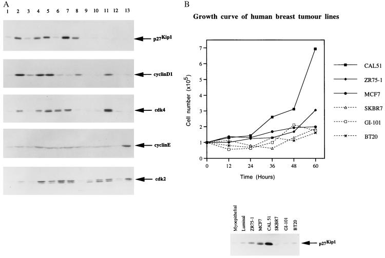
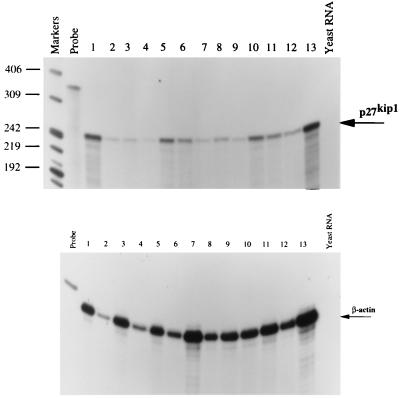
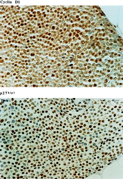
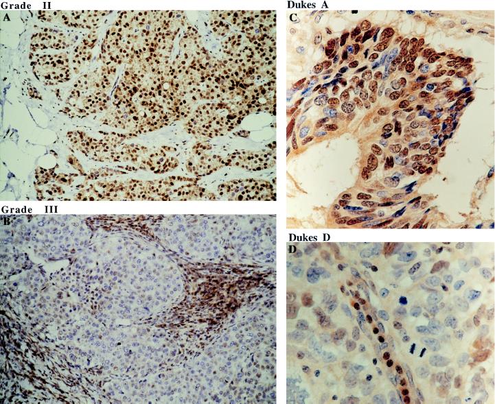
The expression of cyclin-dependent kinase inhibitor p27(kip1) in human tumors and normal tissues was investigated using a panel of novel anti-p27(kip1) mAbs. An inverse correlation between expression of p27(kip1) and cell proliferation was generally observed after analyzing its expression in 25 different normal human tissues. In some highly proliferative human breast cancer cells, however, high level p27(kip1) expression was seen, indicating the existence of a mechanism by which some growing tumor cells may tolerate this inhibitor of cell cycle progression. Detailed studies demonstrated a correlation between the high level expression of p27(kip1) and cyclin D1 in human breast cancer cells. There was also an inverse correlation between the expression of p27(kip1) and the degree of tumor malignancy in human breast and colorectal cancers, indicating that p27(kip1) may be a useful prognostic marker in these cancers.


Figures

Figure 1
Figure 1
Immunoblotting to show the specificity of anti-p27kip1 mAb SX18F7. SX18F7 can specifically recognize the 50-kDa GST-p27kip1 fusion protein (indicated by arrow GST–p27kip1) but not GST. In transfected Saos-2 cells, SX18F7 can specifically react with 27-kDa p27kip1 (indicated by arrow p27kip1) expressed from CMV–p27kip1 plasmid. It does not crossreact with p21waf1/cip1 overexpressed from CMV–p21waf1/cip1 plasmid.
Figure 2
Figure 2
(A) Immunoblotting analysis of p27kip1, cyclin D1, cdk4, cyclin E, and cdk2 expression in human breast cancer cell lines as indicated. The anti-p27kip1 mAb SX53G8 and anti-cyclin D1, anti-cdk4, anti-cyclin E, and anti-cdk2 rabbit polyclonal antibodies are described in Materials and Methods. Lane 1 is the extract from early passaged primary breast myoepithelial cells. Lanes 2–13 are the human breast tumor cell lines: ZR75-1, ZR75-30, MCF-7, MDAMB453, T47D, CAL51, 734B, SKBR5, SKBR7, CAMA, GI-101, and BT20, respectively. (B) Growth curve of six human breast cancer cell lines as indicated (Upper, CAL51, ZR75-1, MCF-7, SKBR7, GI-101, and BT20). (Lower) The p27kip1 protein level of the same group of human breast cancer cell lines is shown. The p27kip1 expression level in the normal primary myoepithelial and luminal epithelial cells is used as a normal control and is labeled as myoepithelial and luminal, respectively.
Figure 3
Figure 3
RNase protection assay to detect the p27kip1 mRNA expression in the human breast cancer cell lines as in Fig. 2. The marker is 32P-labeled MspI-digested pBR322 DNA. The 350-bp p27kip1 RNA probe and the β-actin probe from Ambion are labeled as probe. The loading order of protected p27kip1 mRNA from the 12 breast cancer cell lines are the following. Lanes 1–12 are ZR75-1, SKBR5, SKBR7, MCF-7, MDAMB453, BT20, GI-101, T47D, CAMA, CAL51, ZR75-30, and 734B, respectively. Lane 13 is the protected p27kip1 mRNA from a T lymphoid tumor cell line Jurkat, and the negative control yeast RNA is labeled as indicated. (Lower) The protected β-actin mRNA from the same RNase protection assay is used as a RNA loading control.
Figure 4
Figure 4
Immunohistochemistry staining of cyclin D1 and p27kip1 in the same breast invasive ductal carcinoma using anti-cyclin D1 and anti-p27kip1 mAb DCS-6 and SX53G8 respectively. (×400.)
Figure 5
Figure 5
Immunohistochemistry staining of p27kip1 in human breast and colorectal tumors using anti-p27kip1 mAb SX53G8. (A) Strong nuclear staining of p27kip1 in majority of the tumor cells in grade II invasive ductal carcinoma. (B) In grade III invasive ductal carcinoma, tumor cells are negative for p27kip1, while stromal lymphocytes are strongly stained. (C and D) Nuclear p27kip1 expression is also seen in Dukes A but not Dukes D human colorectal tumors. (A and B, ×250; C and D, ×600.)

References

    1. Draetta G F. Curr. Opin Cell Biol. 1994;6:842–846. - PubMed
    1. Elledge S J, Harper J W. Curr Opin Cell Biol. 1994;6:847–852. - PubMed
    1. Polyak K, Lee M, Erdjument-Bromaga H, Koff A, Roberts J M, Tempst P, Massague J. Cell. 1994;78:59–66. - PubMed
    1. Polyak K, Kato J, Solomon M J, Sherr C J, Massague J, Roberts J M, Koff A. Genes Dev. 1994;8:9–22. - PubMed
    1. Reynisdottir I, Polyak K, Iavarone A, Massague J. Genes Dev. 1995;9:1831–1845. - PubMed

Publication types

MeSH terms