. 1997 Jun 10;94(12):6496-9.
doi: 10.1073/pnas.94.12.6496.

Cleft palate and decreased brain gamma-aminobutyric acid in mice lacking the 67-kDa isoform of glutamic acid decarboxylase

Affiliations

Cleft palate and decreased brain gamma-aminobutyric acid in mice lacking the 67-kDa isoform of glutamic acid decarboxylase

H Asada et al. Proc Natl Acad Sci U S A. .

Abstract

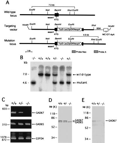
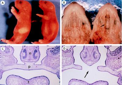
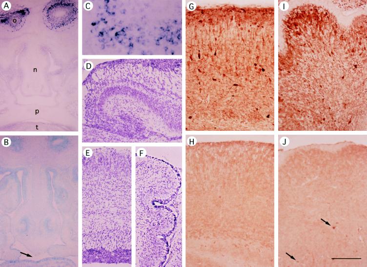
In addition to its role as an inhibitory neurotransmitter, gamma-aminobutyric acid (GABA) is presumed to be involved in the development and plasticity of the nervous system. GABA is synthesized by glutamic acid decarboxylase (GAD), but the respective roles of its two isoforms (GAD65 and 67) have not been determined. The selective elimination of each GAD isoform by gene targeting is expected to clarify these issues. Recently we have produced GAD65 -/- mice and demonstrated that lack of GAD65 does not change brain GABA contents or animal behavior, except for a slight increase in susceptibility to seizures. Here we report the production of GAD67 -/- mice. These mice were born at the expected frequency but died of severe cleft palate during the first morning after birth. GAD activities and GABA contents were reduced to 20% and 7%, respectively, in the cerebral cortex of the newborn GAD67 -/- mice. Their brain, however, did not show any discernible defects. Previous pharmacological and genetic investigations have suggested the involvement of GABA in palate formation, but this is the first demonstration of a role for GAD67-derived GABA in the development of nonneural tissue.


Figures

Figure 1
Figure 1
Targeted disruption of the mouse GAD67 gene. (A) Schematic representation of mouse GAD67 genomic DNA, the targeting vector, and the disrupted gene. A TrpSLacZ/pGKNeo cassette was inserted into the BamHI site of exon 1 located downstream from the translation initiation codon ATG. The location of probes A and Neo that were used for Southern blot analysis are indicated. (B) Southern blot analysis of tail DNA isolated from P0.5 mouse littermates following digestion with EcoRI and probing with the external probe A. (C) RT-PCR analysis of GAD67 (495 bp) and GAD65 transcripts (355 bp) from P0.5 mouse cerebral cortex. RT-PCR of G3PDH mRNA was used to assess the quality of the mRNA. (D and E) Western blot analysis of P0.5 mouse cerebral cortex using anti-GAD65/67 (D) and anti-GAD67 (E) antibodies.
Figure 2
Figure 2
GAD67 knockout mice and cleft palate. (A) Lateral view of wild-type (Left) and GAD67 −/− mutant (Right) newborn mice (P0.5). In homozygous mutants the abdomen is distended but milk is not observed in the stomach. (B) Ventral view of the upper jaw of wild-type (Left) and GAD67 −/− mutant (Right) newborns. Cleft palate (arrows in B and D) was observed in all homozygous mutants. (C and D) Coronal sections of E17.5 facial region of wild-type (C) and GAD67 −/− (D) mice. n, Nasal septa; p, palate; t, tongue.
Figure 3
Figure 3
Expression of GAD67 mRNA and GABA in GAD67 −/− and +/+ brain and head region. (AC) In situ hybridization of frontal sections of E14.5 mouse heads using a GAD67 probe. (A and C) GAD67 +/+. o, olfactory bulb; n, nasal septa; p, palate; t, tongue. C shows the olfactory bulb at higher magnification. (B) GAD67 −/−; counterstained with methyl green. GAD67 signals are found only in neural tissue (olfactory bulb) of GAD67+/+ or +/− mice. (DF) Hematoxylin-eosin staining of P0.5 GAD67 −/− brain. (D) Hippocampus. (E) Cerebral cortex. (F) Cerebellum. (GJ) Immunohistochemistry with anti-GABA antibody (Sigma). (G and H) Cerebral cortex. (I and J) Cerebellum. (G and I) P0.5 wild-type mice. (H and J) P0.5 null mutant mice. Arrows in J indicate GABA-positive neurons in the deep part of GAD67 −/− cerebellum. (Bar in J = 500 μm for A, B, and DF; 200 μm for GJ; and 100 μm for C.)

References

    1. Obata K. Int Rev Neurobiol. 1972;15:167–187. - PubMed
    1. Roberts E, Chase T N, Tower D B, editors. GABA in Nervous System Function. New York: Raven; 1976.
    1. Erlander M G, Tobin A J. Neurochem Res. 1991;16:215–226. - PubMed
    1. Erlander M G, Tillakaratne N J K, Feldblum S, Patel N, Tobin A J. Neuron. 1991;7:91–100. - PubMed
    1. Ma W, Chang L, Zhang L, Barker J L. J Neurosci. 1995;15:2547–2560. - PMC - PubMed

Publication types

MeSH terms