. 1997 Jun 10;94(12):6529-34.
doi: 10.1073/pnas.94.12.6529.

Elevation of intracellular calcium in smooth muscle causes endothelial cell generation of NO in arterioles

Affiliations

Elevation of intracellular calcium in smooth muscle causes endothelial cell generation of NO in arterioles

K A Dora et al. Proc Natl Acad Sci U S A. .

Abstract

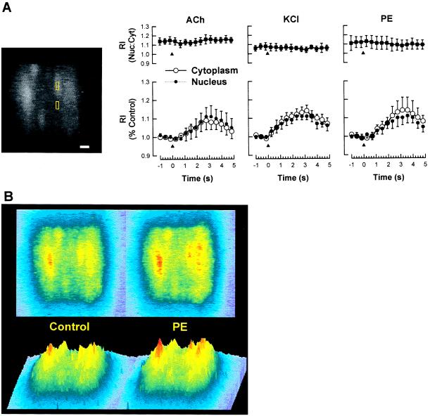
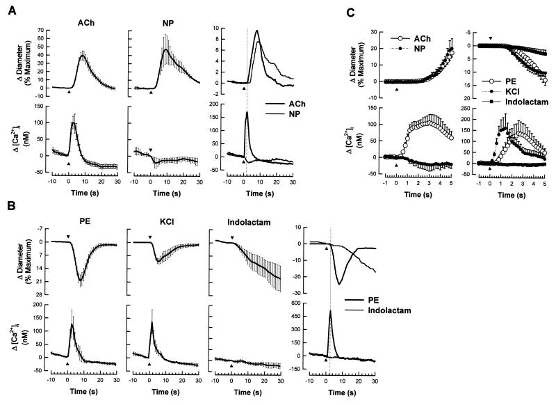
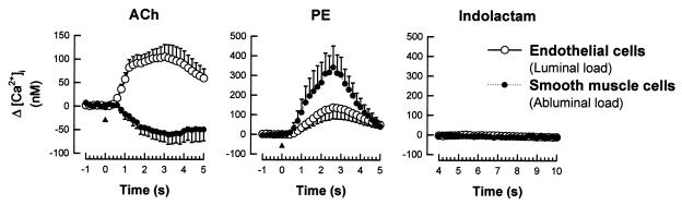
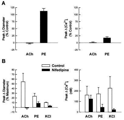
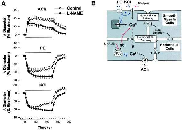
It is well known that vascular smooth muscle tone can be modulated by signals arising in the endothelium (e.g., endothelium-derived relaxing factor, endothelium-derived hyperpolarizing factor, and prostaglandins). Here we show that during vasoconstriction a signal can originate in smooth muscle cells and act on the endothelium to cause synthesis of endothelium-derived relaxing factor. We studied responses to two vasoconstrictors (phenylephrine and KCl) that act by initiating a rise in smooth muscle cell intracellular Ca2+ concentration ([Ca2+]i) and exert little or no direct effect on the endothelium. Fluo-3 was used as a Ca2+ indicator in either smooth muscle or endothelial cells of arterioles from the hamster cheek pouch. Phenylephrine and KCl caused the expected rise in smooth muscle cell [Ca2+]i that was accompanied by an elevation in endothelial cell [Ca2+]i. The rise in endothelial cell [Ca2+]i was followed by increased synthesis of NO, as evidenced by an enhancement of the vasoconstriction induced by both agents after blockade of NO synthesis. The molecule involved in signal transmission from smooth muscle to endothelium is as yet unknown. However, given that myoendothelial cell junctions are frequent in these vessels, we hypothesize that the rise in smooth muscle cell Ca2+ generates a diffusion gradient that drives Ca2+ through myoendothelial cell junctions and into the endothelial cells, thereby initiating the synthesis of NO.


Figures

Figure 3
Figure 3
Image analysis of arteriolar endothelial cell [Ca2+]i responses to agonists. In these experiments the plane of focus was lowered to the layer of endothelial cells lining the bottom surface of the arteriole (hence no diameter responses were obtained). (A) Analysis of recorded video images allowed determination of nuclear and cytosolic changes in fluorescence intensity. (Left) Endothelial cells are orientated longitudinally along the arteriole (top to bottom) and are readily distinguished from smooth muscle cells (not visible). Small boxes were positioned in the two compartments (three cells per image) and relative intensity (RI) over time was recorded. Note: In all experiments only a short segment of arteriole was exposed to epi-illumination. (Bar = 10 μm.) (Right) Each agonist increased both nuclear (Nuc) and cytosolic (Cyt) fluorescence intensity in equal proportions. ACh (n = 6), PE (n = 4), and KCl (n = 4) were applied at the same concentration and for the same duration as described in Fig. 1. After a response to ACh was obtained, PE and/or KCl was applied at the same arteriolar location. Values are means ± SE; ▴ denotes time at which agonist application commenced. (B) Pseudocolor images of arteriolar endothelial cells before (Control) and 1 sec after (PE) stimulation with 10−4 M PE. Bottom panels are profiles of the corresponding top panel images.
Figure 1
Figure 1
Time course of changes in vessel diameter and endothelial cell [Ca2+] in response to abluminal pressure-pulse application of agonists to isolated perfused resistance arterioles. (A) ACh (10−4 M, 1.2-sec pulse, n = 15) and sodium nitroprusside (NP 5 × 10⁻⁵ M, 1.2-sec pulse, n = 3) both stimulated vasodilation, but had opposite effects on [Ca2+]i. (Right) The rapid rise in [Ca2+]i in response to ACh, but not sodium nitroprusside, at the same arteriolar location suggests changes in fluorescence intensity relate to changes in endothelial cell rather than smooth muscle [Ca2+]i (typical trace). (B) PE (10−5 M, 1.2-sec pulse, n = 11) and KCl (250 mM, 2.2-sec pulse, n = 8) both stimulated vasoconstriction and rises in endothelial cell [Ca2+]i. In contrast, indolactam (10−5 M, 1.2-sec pulse, n = 4) caused vasoconstriction without a change in [Ca2+]i. (Right) No increase in [Ca2+]i was observed in association with indolactam-stimulated vasoconstriction at the same arteriolar location shown to be responsive to PE (typical trace). (C) For each agonist, changes in endothelial cell [Ca2+]i occurred well in advance of changes in arteriolar diameter. Values are means ± SE; ▴ denotes time at which agonist application commenced.
Figure 2
Figure 2
Comparison between smooth muscle and endothelial cell [Ca2+]i responses to abluminal pressure-pulse application of agonists. Data from Fig. 1 are compared with responses obtained after loading smooth muscle cells with fluo-3 by adding the dye to the superfusion medium (abluminal load) rather than perfusion medium (luminal load). ACh (n = 5), PE (n = 6), and indolactam (n = 4) were applied at the same concentration and for the same duration as described in Fig. 1. Peak diameter responses were the same between groups. Values are means ± SE; ▴ denotes time at which agonist application commenced.
Figure 4
Figure 4
(A) Effect of endothelium-specific damage on the diameter and fluorescence responses to agonists. Perfusion of air bubbles through the lumen of arterioles changed resting diameter from 64.9 ± 4.9 to 60.5 ± 10.5 μm (n = 4). The responses to ACh were abolished (n = 4), whereas PE-mediated vasoconstriction was unaffected, yet the increase in fluorescence was markedly attenuated (n = 4). (B) Effect of nifedipine on changes in vessel diameter and endothelial cell [Ca2+] in response to agonists. Addition of nifedipine (10−6 M) to the superfusion solution resulted in near maximal dilation of the arteriole (Δ diameter 64 ± 4 to 84 ± 5 μm, n = 8). In the presence of nifedipine, the rise in [Ca2+]i in response to ACh was not affected (106.3 ± 27.6% of control) despite an absence of vasodilation (−8.1 ± 8.1% of control, n = 5). In contrast, both the rise in [Ca2+]i and vasoconstriction in response to PE were reduced (30.9 ± 14.8%, 37.1 ± 9.6% of control, respectively, n = 4) and were markedly reduced in response to KCl (23.4 ± 9.2%, 25.7 ± 10.4% of control, respectively, n = 5). For all agents, values are means ± SE of paired responses before and after each treatment. The stimulation pipette was positioned at the same arteriolar location to obtain each pair of responses.
Figure 5
Figure 5
(A) Effect of l-NAME on the time course of changes in vessel diameter in response to agonists. ACh (10−4 M, n = 5), PE (10−5 M, n = 5), and KCl (250 mM, n = 5) were pressure-pulse ejected adjacent to a segment of the arteriole for a period of 2 min (▪). l-NAME (10−5 M) was added to the superfusion solution and allowed to equilibrate for 20 min, and had no effect on vessel diameter (Δ Diameter from 64.5 ± 3.4 to 66.8 ± 3.4 μm, n = 15). Without changing the stimulation pipette position, the time course for each agonist was repeated in the presence of l-NAME. Values are means ± SE of paired responses before and after l-NAME. (B) Schematic of the proposed heterocellular Ca2+ diffusion pathway. Increases in smooth muscle [Ca2+]i stimulated by either PE or KCl diffuse radially to underlying endothelial cells through myoendothelial gap junctions. The secondary increase in endothelial cell [Ca2+]i stimulates NO synthase to generate NO and modulate smooth muscle [Ca2+]i and hence contraction. The extent of homocellular Ca2+ diffusion is limited by cell volume and distance.

References

    1. Busse, R., Pohl, U. & Luckhoff, A. (1989) Z. Kardiol. 78, Suppl. 6, 64–69. - PubMed
    1. von der Weid P Y, Beny J L. Am J Physiol. 1992;262:H1823–H1831. - PubMed
    1. Angus J A, Cocks T M, Satoh K. Fed Proc Fed Am Soc Exp Biol. 1986;45:2355–2359. - PubMed
    1. Amerini S, Mantelli L, Ledda F. Pharmacol Res. 1995;31:175–181. - PubMed
    1. Ayajiki K, Kindermann M, Hecker M, Fleming I, Busse R. Circ Res. 1996;78:750–758. - PubMed

Publication types

MeSH terms

LinkOut - more resources