. 1997 Jun 16;137(6):1199-210.
doi: 10.1083/jcb.137.6.1199.

Integral membrane proteins of the nuclear envelope are dispersed throughout the endoplasmic reticulum during mitosis

Affiliations

Integral membrane proteins of the nuclear envelope are dispersed throughout the endoplasmic reticulum during mitosis

L Yang et al. J Cell Biol. .

Abstract

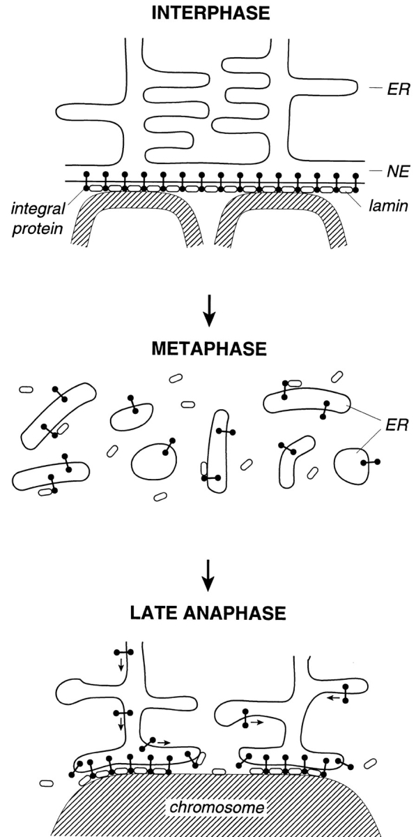
We have analyzed the fate of several integral membrane proteins of the nuclear envelope during mitosis in cultured mammalian cells to determine whether nuclear membrane proteins are present in a vesicle population distinct from bulk ER membranes after mitotic nuclear envelope disassembly or are dispersed throughout the ER. Using immunofluorescence staining and confocal microscopy, we compared the localization of two inner nuclear membrane proteins (laminaassociated polypeptides 1 and 2 [LAP1 and LAP2]) and a nuclear pore membrane protein (gp210) to the distribution of bulk ER membranes, which was determined with lipid dyes (DiOC6 and R6) and polyclonal antibodies. We found that at the resolution of this technique, the three nuclear envelope markers become completely dispersed throughout ER membranes during mitosis. In agreement with these results, we detected LAP1 in most membranes containing ER markers by immunogold electron microscopy of metaphase cells. Together, these findings indicate that nuclear membranes lose their identity as a subcompartment of the ER during mitosis. We found that nuclear lamins begin to reassemble around chromosomes at the end of mitosis at the same time as LAP1 and LAP2 and propose that reassembly of the nuclear envelope at the end of mitosis involves sorting of integral membrane proteins to chromosome surfaces by binding interactions with lamins and chromatin.


Figures

Figure 1
Figure 1
Localization of ER membranes with antibodies and fluorescent dyes. NRK cells were fixed and labeled for indirect immunofluorescence microscopy with a polyspecific anti-ER antibody (A and B) or anti–protein disulphide isomerase antibody (anti-PDI, C and D) and were then counterstained with DiOC6. Alternatively, the fixed cells were double stained with DiOC6 and R6 (E and F). Shown are images of the labeling with DiOC6 (left column), antibodies or R6 (center column), and the merge of the two fluorescent channels (right column). Examples of interphase (A, C, and E) and metaphase (B, D, and F) are presented. Bar, 10 μm.
Figure 2
Figure 2
Comparison of the localizations of LAP1 and ER membranes throughout mitosis. NRK cultures enriched in mitotic cells were fixed and examined by double immunofluorescence microscopy after labeling with the LAP1-specific monoclonal antibody RL13 and a polyspecific anti-ER antibody. Shown is the labeling with RL13 (left column), the anti-ER antibody (center column), and the merge of the two fluorescent images (right column). The figure presents representative examples of cells (two different cells in each row) in interphase (A), prophase (B, left cell), prometaphase (B, right cell), metaphase (C), mid-anaphase (D, left cell), late anaphase (D, right cell) and telophase (E). The NE in the prophase cell shown in B (left cell) is deformed due to invagination by the mitotic spindle. Bar, 10 μm.
Figure 3
Figure 3
Comparison of the localization of ER membranes to the distributions of LAP2, gp210, and α-mannosidase II in metaphase cells. NRK cultures enriched in mitotic cells were fixed and examined by double labeling with DiOC6 and the LAP2-specific monoclonal antibody RL29 (A), DiOC6 and the gp210-specific monoclonal antibody RL20 (B), or R6 and a polyclonal antibody against the Golgi enzyme α-mannosidase II (C). Shown is the labeling with the ER probes (left column), antibody probes (center column), and a merge of the two fluorescent images (right column). The green/red color representation for the antibody and R6 was computationally reversed in the images presented in C for consistency. In A and B, two different metaphase cells are shown, and in C, a metaphase (left cell) and anaphase (right cell) are shown. Bar, 10 μm.
Figure 4
Figure 4
Comparison of the localizations of LAP1 and ER membranes in metaphase cells by double immunogold labeling. Populations of nocodazole-arrested metaphase NRK cells were processed for double immunogold labeling by incubation with the LAP1-specific monoclonal antibody RL13 and a polyspecific anti-ER antibody, followed by 10-nm antibody-coupled gold (to detect the LAP1 probe) and 5-nm antibody-coupled gold (to detect the ER probe). Shown are images of cells incubated with anti-LAP1 and anti-ER antibodies followed by secondary gold-coupled antibodies (A and C) or cells incubated with only the secondary gold-coupled antibodies and without the primary antibodies (B). Examples of mitotic chromosomes (ch) used to identify mitotic cells are designated. A indicates examples of the two classes of antibody-labeled structures: large discrete vesicles (large arrowheads) and densely staining aggregates of small vesicles and tubules (small arrows). C shows a gallery of the antibody-labeled membranes: large discrete vesicles (top row) and aggregates of small vesicles and tubules (bottom two rows). Bars: (A and B) 300 nm; (C) 100 nm.
Figure 5
Figure 5
Comparison of the localization of lamins to the distribution of LAPs at the end of mitosis. NRK cultures enriched in mitotic cells were labeled for double immunofluorescence microscopy with an antibody against lamin A and the LAP1-specific monoclonal antibody RL13 (A and B) or the LAP2-specific monoclonal antibody RL29 (C and D). Shown are images of the lamin labeling (left column), the LAP1 or LAP2 labeling (center column), or a merge of the two fluorescent images (right column). Each row presents representative images of cells in late anaphase (left cell) or mid-late telophase (right cell). Bar, 15 μm.
Figure 6
Figure 6
Model depicting the dynamics of integral membrane proteins of the NE during mitosis. The interphase NE is morphologically continuous with the peripheral ER but is a specialized ER subcompartment that contains integral membrane proteins specific to the inner nuclear membrane (top) and nuclear pore membrane (not shown). Integral proteins of the inner nuclear membrane may be bound to lamins, lamina-associated proteins, or chromatin. During mitosis when the NE is disassembled, integral membrane proteins of the NE are dispersed throughout all ER membranes, and the NE loses its identity as an ER subcompartment (middle). We propose that integral proteins are resorted to the NE during late anaphase by diffusion through a functionally continuous ER and subsequent association with binding sites at the chromosomes (bottom). The binding sites for inner membrane proteins may include lamins and chromatin (bottom); those for nuclear pore membrane proteins may include other pore complex components (not shown). These binding interactions would result in the net collection of nuclear membrane proteins at the reforming NE (bottom, arrows). Reformation of the NE may involve cooperative assembly of lamins and integral membrane proteins of the inner nuclear membrane (bottom).

References

    1. Burke B. On the cell-free association of lamins A and C with metaphase chromosomes. Exp Cell Res. 1990;186:169–176. - PubMed
    1. Burke B, Gerace L. A cell-free system to study reassembly of the nuclear envelope at the end of mitosis. Cell. 1986;44:639–652. - PubMed
    1. Chaudhary N, Courvalin JC. Stepwise reassembly of the nuclear envelope at the end of mitosis. J Cell Biol. 1993;122:295–306. - PMC - PubMed
    1. Foisner R, Gerace L. Integral membrane proteins of the nuclear envelope interact with lamins and chromosomes, and binding is modulated by mitotic phosphorylation. Cell. 1993;73:1267–1279. - PubMed
    1. Furukawa K, Pante N, Aebi U, Gerace L. Cloning of a cDNA for lamina-associated polypeptide 2 (LAP2) and identification of regions that specify targeting to the nuclear envelope. EMBO (Eur Mol Biol Organ) J. 1995;14:1626–1636. - PMC - PubMed

Publication types

MeSH terms