. 1997 Jun 16;137(6):1287-307.
doi: 10.1083/jcb.137.6.1287.

Unconventional myosins in inner-ear sensory epithelia

Affiliations

Unconventional myosins in inner-ear sensory epithelia

T Hasson et al. J Cell Biol. .

Abstract

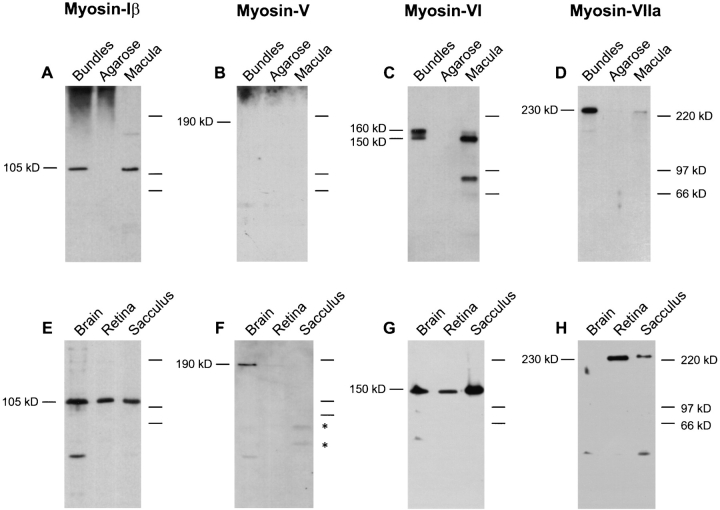
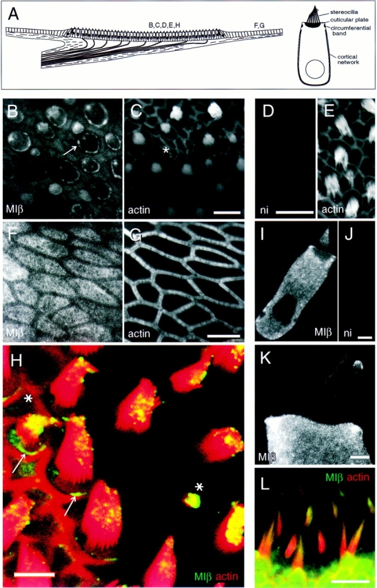
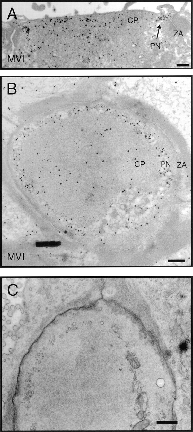
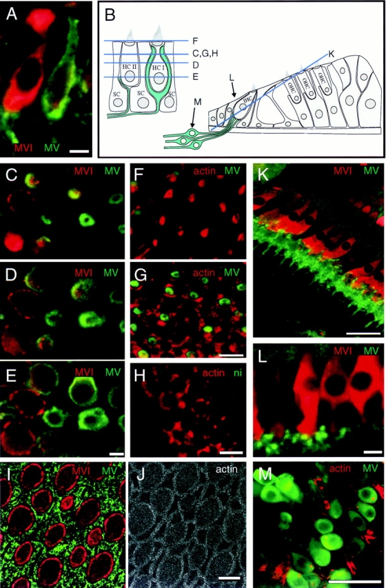
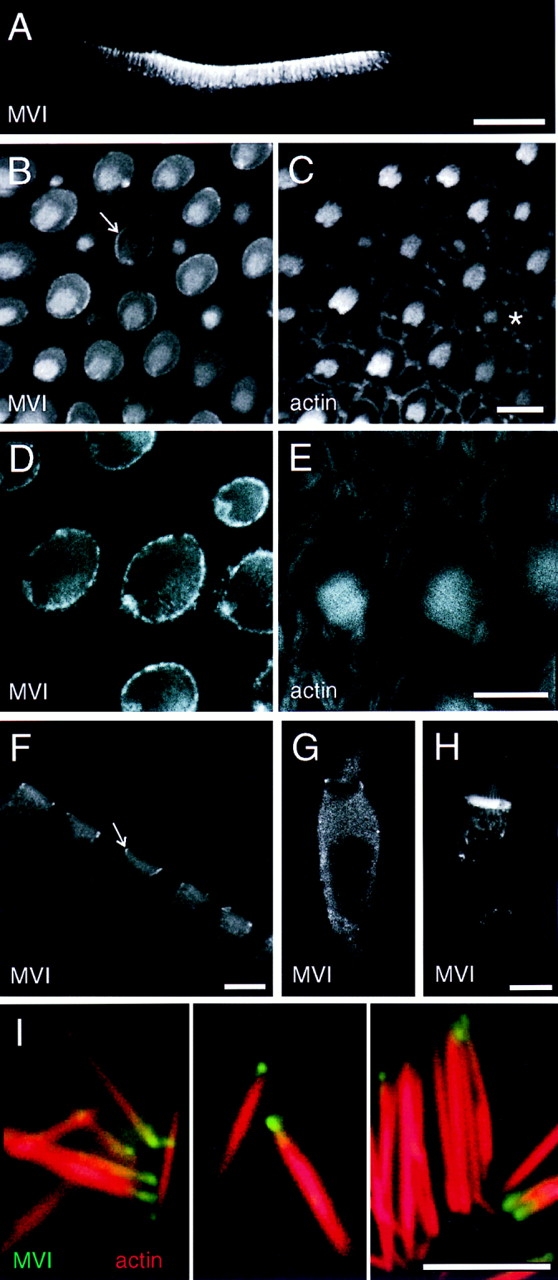
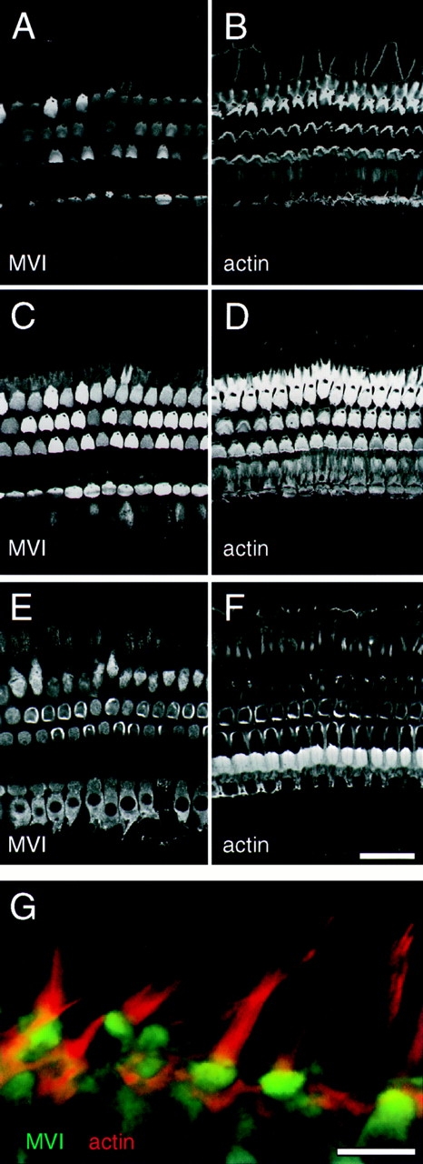
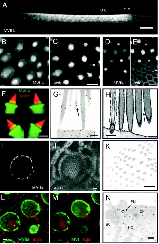
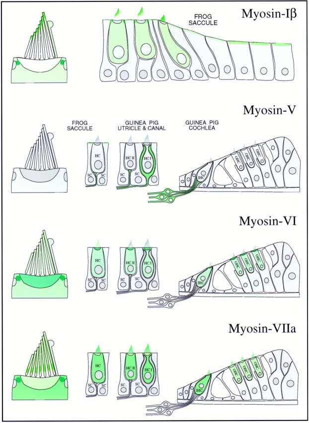
To understand how cells differentially use the dozens of myosin isozymes present in each genome, we examined the distribution of four unconventional myosin isozymes in the inner ear, a tissue that is particularly reliant on actin-rich structures and unconventional myosin isozymes. Of the four isozymes, each from a different class, three are expressed in the hair cells of amphibia and mammals. In stereocilia, constructed of cross-linked F-actin filaments, myosin-Ibeta is found mostly near stereociliary tips, myosin-VI is largely absent, and myosin-VIIa colocalizes with crosslinks that connect adjacent stereocilia. In the cuticular plate, a meshwork of actin filaments, myosin-Ibeta is excluded, myosin-VI is concentrated, and modest amounts of myosin-VIIa are present. These three myosin isozymes are excluded from other actin-rich domains, including the circumferential actin belt and the cortical actin network. A member of a fourth class, myosin-V, is not expressed in hair cells but is present at high levels in afferent nerve cells that innervate hair cells. Substantial amounts of myosins-Ibeta, -VI, and -VIIa are located in a pericuticular necklace that is largely free of F-actin, squeezed between (but not associated with) actin of the cuticular plate and the circumferential belt. Our localization results suggest specific functions for three hair-cell myosin isozymes. As suggested previously, myosin-Ibeta probably plays a role in adaptation; concentration of myosin-VI in cuticular plates and association with stereociliary rootlets suggest that this isozyme participates in rigidly anchoring stereocilia; and finally, colocalization with cross-links between adjacent stereocilia indicates that myosin-VIIa is required for the structural integrity of hair bundles.


Figures

Figure 1
Figure 1
Protein immunoblot detection of unconventional myosin isozymes expressed in frog hair bundles and tissues. (Top panels) Frog saccular hair bundles were isolated by the twist-off method (Gillespie and Hudspeth, 1991). Bundles, ∼40,000 hair bundles (21 saccular equivalents). Agarose, ∼2 mg of agarose, from agarose adjacent to purified bundles but free of tissue, as a control. Macula, sensory epithelia cells (without peripheral cells, basement membrane, or nerve) remaining after bundle isolation. Protein for ∼1.0 sensory epithelium (2,000 hair cells and 4,000 supporting cells) was loaded. Proteins were separated by SDS-PAGE, transferred to PVDF membranes, and probed with antibodies specific for myosin-Iβ (A and E), -V (B and F), -VI (C and G), and -VIIa (D and H), as described in the text. (Bottom panels) Total protein (10 μg) from brain, retina, and whole saccule was loaded. On low cross-linker gels such as these, myosin-Iβ migrates with an estimated molecular mass of ∼105 kD. Asterisks in F indicate saccular proteins that cross-react with the 32A antibody. Detection was with the following antibodies: (A and E) rafMIβ; (B and F) 32A; (C and G) rapMVI; (D and H) rahMVIIa.
Figure 2
Figure 2
Localization of myosin-Iβ. (A, left) Depiction of a vertical cross-section through a frog saccular epithelium. In the sensory epithelium, the central region in this illustration, ∼2,000 hair cells and ∼4,000 supporting cells are packed in a regular array. Afferent and efferent nerve fibers penetrate a basement membrane before contacting hair cells on their basolateral surfaces. Outside the sensory epithelium, peripheral cells are arranged in a simple cuboidal epithelium. Letters indicate viewpoints of subsequent panels. (Right) Depiction of a single saccular hair cell, showing actin-rich domains. (B and C) Frog saccule hair cells labeled for myosin-Iβ in B and actin in C. Optical section at apical surface at low magnification. Note strong pericuticular necklace labeling (arrow in B), lesser labeling within cuticular plates, and bright labeling of small bundles (asterisk in C). Also note lack of staining in junctional actin bands. (D and E) Frog saccule hair cells labeled with nonimmune control antibody in D; corresponding actin labeling in E. (F and G) Labeling for myosin-Iβ in frog saccule peripheral cell region in F; corresponding actin labeling in G. Apical surfaces are labeled well with myosin-Iβ antibody, except where circumferential actin belts are present. (H) High magnification view of frog saccular hair bundles labeled for myosin-Iβ (green) and actin (red). Colocalization (yellow) is strongest at stereociliary tips. (Asterisks) Small hair bundles, which are strongly labeled; (arrow) the pericuticular necklace, clearly segregated from actin of the cuticular plate and of the circumferential actin belt. Single optical section. (I) Myosin-Iβ in single dissociated frog saccule hair cell. Label is excluded from the nucleus and cuticular plate. Single optical section. (J) Single dissociated cell labeled with irrelevant affinity-purified antibody. (K) High magnification view of a single dissociated frog saccule hair cell labeled for myosin-Iβ. Note preferential labeling of stereocilia tips and labeling of kinociliary bulbs. Projection of 12 optical sections across middle third of bundle. (L) Labeling of rat utriculus with mAb 20-3-2 (green), raised against bovine myosin-Iβ; actin is shown in red. Note intense myosin-Iβ reactivity near tips of stereocilia. Bars: (B and C) 10 μm; (D and E) 25 μm; (F and G) 5 μm; (H) 10 μm; (I and J) 5 μm; (K) 2 μm; (L) 10 μm.
Figure 3
Figure 3
Localization of myosin-Iβ in frog saccule by immunoelectron microscopy. (A) Immunoelectron microscopy with rafMIβ and protein A–gold detection showing labeling at stereociliary insertions. Myosin-Iβ is particularly enriched at the rootlet density (arrow). (B) Near-horizontal cross-section through the same region as shown in A, passing from cuticular plate (bottom) to bases of stereocilia (top). (Inset) The plane of section. Label appears where stereocilia join the cuticular plate (arrows) but not above (arrowhead). (C) Gold labeling at pericuticular necklace. SC, supporting cell; HC, hair cell. The hair cell/supporting cell junction is marked by the electron-dense band. (D) Gold labeling at upper end of stereocilia. Bars: (A–C) 1 μm; (D) 500 nm.
Figure 6
Figure 6
Immunoelectron microscopic localization of myosin-VI in frog saccule. (A) Vertical cross-section through the cuticular plate region showing pericuticular necklace labeling (PN) between cuticular plate (CP) and circumferential actin belt at the zonula adherens (ZA). (B) Horizontal section through the cuticular plate and zonula adherens. Label in the hair cell at this level is strongest in the regions not occupied by actin. (C) Same level as B but with more rapid fixation and without antibody labeling with its extensive tissue extraction. Cytoplasmic vesicles are visible in the pericuticular necklace region. Bars: (A–C) 1 μm.
Figure 4
Figure 4
Localization of myosin-V in guinea pig utricle and cochlea and in frog saccule. All images are single optical sections. (A) Double labeling showing myosin-V (green) in neuronal processes contacting type II (left) and type I (right) hair cells in guinea pig utricule; hair cells also labeled for myosin-VI (red) to visualize cell bodies. (B) Depiction of guinea pig utricule on left and cochlea on right. Optical section levels for some images are indicated. (C–E) Confocal z-series through utricular hair cells labeled for myosin-V (green) and myosin-VI (red), showing rings of myosin-V labeling associated with calyces enveloping type I hair cells. (F and G) Labeling of utricular cells for myosin-V (green) and actin (red), showing myosin-V staining is absent from stereocilia in F and from circumferential actin belts in G. (H) Nonimmune control labeling (green) of utricular hair cells at the level of the circumferential actin belt; actin labeling is shown in red. (I and J) Apical surface of bullfrog saccule, triple labeled for myosin-V (green, I), myosin-VI (red, I), and actin (J); myosin-V labeling is associated with supporting cell apical surfaces. (K) Section through guinea pig cochlea, stained for myosin-V (green) and -VI (red). Myosin-V labeling is associated with neuronal processes contacting the bases inner hair cells. (L) High magnification view of bouton endings contacting cochlear inner hair cells; stained for myosin-V (green) and -VI (red). (M) Spiral ganglion neurons stained for myosin-V (green) and actin (red). Bars: (A, C–J, and L) 10 μm; (K and M) 50 μm.
Figure 5
Figure 5
Localization of myosin-VI in frog saccule. (A) Confocal image of Vibratome section of saccular epithelium at low magnification, labeled for myosin-VI. Myosin-VI is found nearly exclusively in hair cells. (B and C) Sections through apical surface of saccular sensory epithelium showing myosin-VI in B and actin in C. Note strong pericuticular necklace labeling (arrow in B) and bright labeling of small bundles (asterisk in C). (D and E) High magnification apical section showing myosin-VI labeling in D and actin labeling in E. Note strong myosin-VI labeling of the pericuticular necklace; actin is excluded from this structure. (F) Vibratome section showing cuticular plate labeling in hair cells. The pericuticular necklace is also visible in cross-section (arrow). (G) Myosin-VI labeling in a dissociated hair cell, fixed before antibody incubation. (H) Myosin-VI labeling in a dissociated hair cell, permeabilized with streptolysin O and incubated with myosin-VI antibody overnight before fixation. Hair cells prepared in this manner had very strong cuticular plate immunoreactivity. (I) High power views of isolated stereocilia labeled for myosin-VI (green) and actin (red). Myosin-VI is enriched at the tapered end of the stereocilia. Bars: (A) 100 μm; (B–H) 10 μm; (I) 5 μm.
Figure 7
Figure 7
Localization of myosin-VI in guinea pig auditory and vestibular epithelia. (A–F) Labeling of cochlear hair cells for myosin-VI (A, C, and E) and actin (B, D, and F). Three successive optical sections through the organ of Corti, the sensory epithelium of the cochlea. (A and B) Optical section at the level of the stereocilia (0 μm). Hair bundles are V-shaped in outer hair cells (top three rows), and straight in inner hair cells (bottom row). Myosin-VI is not present in these cochlear stereocilia. (C and D) Optical section at −1.4 μm, at the level of the cuticular plates. Myosin-VI is enriched at this level. (E and F) Optical section at −4.3 μm, at the level of cell bodies of the inner and outer hair cells. Myosin-VI is present throughout cochlear hair cell bodies. (G) Side view of utricular hair cells, labeled for myosin-VI (green) and actin (red). No label is present in stereocilia. Bars: (A–F) 50 μm; (G) 10 μm.
Figure 8
Figure 8
Localization of myosin-VIIa in frog saccule. (A) Vibratome section of saccular epithelium at low magnification, labeled for myosin-VIIa. Myosin-VIIa is found nearly exclusively in hair cells. Positions of some images are indicated. (B and C) Vertical view of the middle of sensory epithelium labeled for myosin-VIIa in B and actin in C. Myosin-VIIa is present in stereocilia and the pericuticular necklace; small bundles are also intensely labeled (asterisk in C). (D and E) Vertical view of the edge of sensory epithelium (periphery is on bottom) labeled for myosin-VIIa in D and actin in E. Note small bundles are intensely labeled for myosin-VIIa (asterisk). (F) Four isolated hair cells, labeled from myosin-VIIa (green) and actin (red). The yellow bands toward the bases of stereocilia indicate particularly high concentrations of myosin-VIIa. (G) Immunoelectron microscopy showing concentration of myosin-VIIa (arrow) in a band immediately above basal tapers. (H) Electron micrograph of unlabeled tissue showing ankle links in the same region (arrow) as label in G. (I and J) High resolution view of one hair cell, showing concentration of myosin-VIIa label in the pericuticular necklace. Note in I the punctate nature of myosin-VIIa labeling in the pericuticular necklace, and its separation from the actin domains seen in J. (K) Immunoelectron microscopy cross-section through a hair bundle, with the plane of section passing from insertions (lower left) to above the tapers (upper right). Myosin-VIIa label occurs only above taper region. (L and M) Triple-labeling comparison of myosin-VIIa, myosinVI, and actin in the same sample. In L, myosin-VIIa (green); actin (red). In M, myosin-VI (green); actin (red). Note that the pattern of myosin-VIIa and -VI labeling in the pericuticular necklace is very similar in most cells. (N) Immunoelectron microscopy showing myosin-VIIa in pericuticular necklace (PN) and cuticular plate (CP). Hair cell (HC) and supporting cell (SC) are also indicated. Bars: (A) 100 μm; (B–F) 10 μm; (G and H) 500 nm; (I, J, L, and M) 2 μm; (K and N) 1 μm.
Figure 9
Figure 9
Localization of myosin-VIIa in mammalian cochlea, utricule, and semicircular canal. (A) Labeling of mouse cochlear hair cells labeled for myosin-VIIa (green) and actin (red). This optical section is slightly askew, revealing both hair bundles and cell bodies. Note apparently uniform myosin-VIIa labeling in hair bundles. (B and C) Hair bundles of mouse utricle, labeled for myosin-VIIa in B and actin in C. (D and E) Guinea pig semicircular canal hair cells, labeled for myosin-VIIa in D and actin in E. Note that myosin-VIIa is in both type I and type II hair cells, and throughout the long stereocilia. Bars: (A–E) 10 μm.
Figure 10
Figure 10
Myosin isozyme location in sensory epithelia of auditory and vestibular organs. Green shading indicates myosin location; darker green indicates higher myosin concentration. (Myosin-Iβ) The strongest labeling is in the pericuticular necklace; modest amounts of myosin-Iβ are found in the hair cell cytoplasm and apical surfaces of peripheral cells. Within the bundle, myosin-Iβ labeling is focused toward stereociliary tips. (Myosin-V) No labeling in hair cells; all labeling in sensory epithelium apparently is associated with afferent nerve fibers. Myosin-V is found in both calyx and bouton synaptic terminals. (Myosin-VI) Hair cells are strongly labeled; supporting cells are not labeled at all. The highest concentration is in cuticular plates and pericuticular necklaces. There is light hair bundle labeling in frog but not in guinea pig. (Myosin-VIIa) Myosin-VIIa is expressed exclusively in hair cells, throughout the cell bodies and along the lengths of stereocilia. In frog, myosin-VIIa is found throughout stereocilia but most strikingly in a band immediately above the basal connectors (also called ankle links); substantial amounts are also found in the pericuticular necklace.

References

    1. Assad JA, Corey DP. An active motor model for adaptation by vertebrate hair cells. J Neurosci. 1992;12:3291–3309. - PMC - PubMed
    1. Avraham KB, Hasson T, Steel KP, Kingsley DM, Russell LB, Mooseker MS, Copeland NG, Jenkins NA. The mouse Snell's waltzer deafness gene encodes an unconventional myosin required for structural integrity of inner ear hair cells. Nat Genet. 1995;11:369–375. - PubMed
    1. Bagshaw, C.R. 1993. Muscle Contraction. Second edition. Chapman and Hall, London. 155 pp.
    1. Baines IC, Brzeska H, Korn ED. Differential localization of Acanthamoebamyosin I isoforms. J Cell Biol. 1992;5:1193–1203. - PMC - PubMed
    1. Baines IC, Corigliano-Murphy A, Korn ED. Quantification and localization of phosphorylated myosin I isoforms in Acanthamoeba castellanii. . J Cell Biol. 1995;130:591–603. - PMC - PubMed

Publication types