. 1997 Jun 16;137(6):1321-36.
doi: 10.1083/jcb.137.6.1321.

Spindle dynamics during meiosis in Drosophila oocytes

Affiliations

Spindle dynamics during meiosis in Drosophila oocytes

S A Endow et al. J Cell Biol. .

Abstract

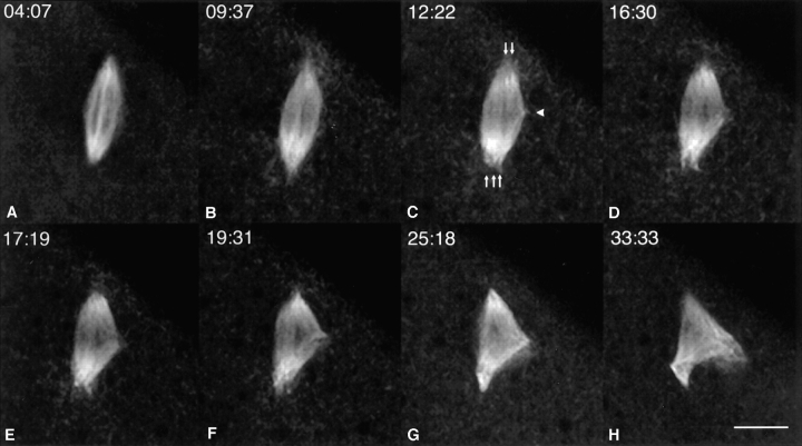
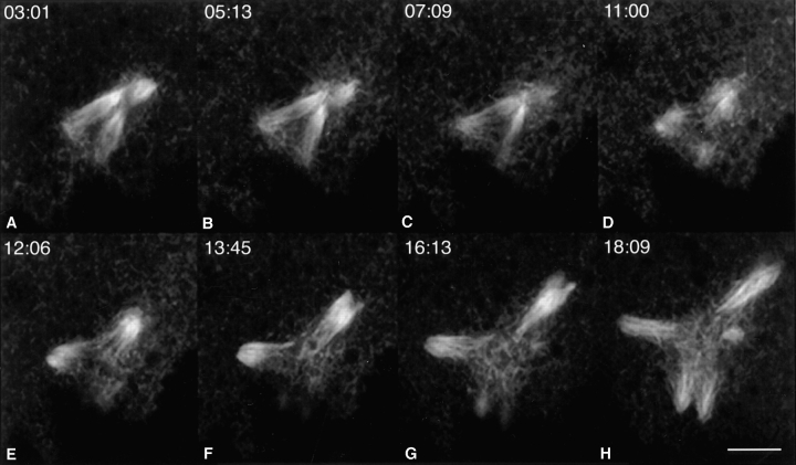
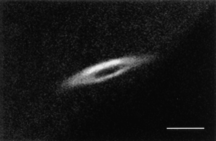
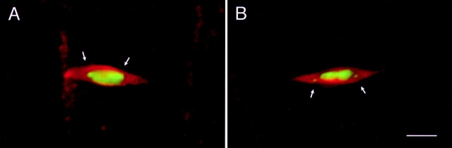
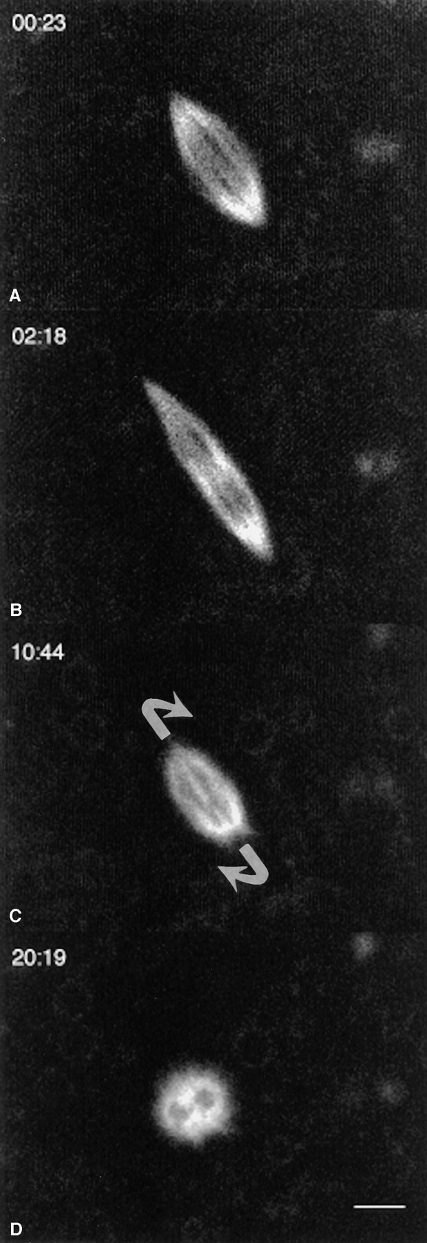
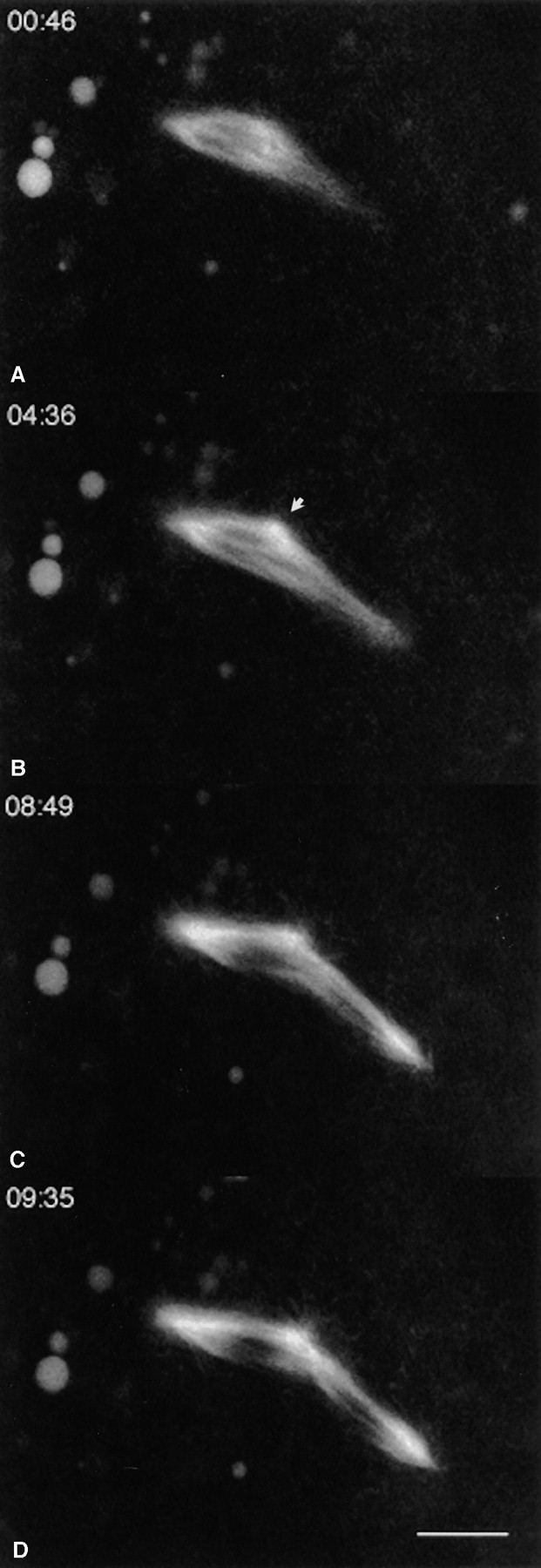
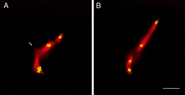
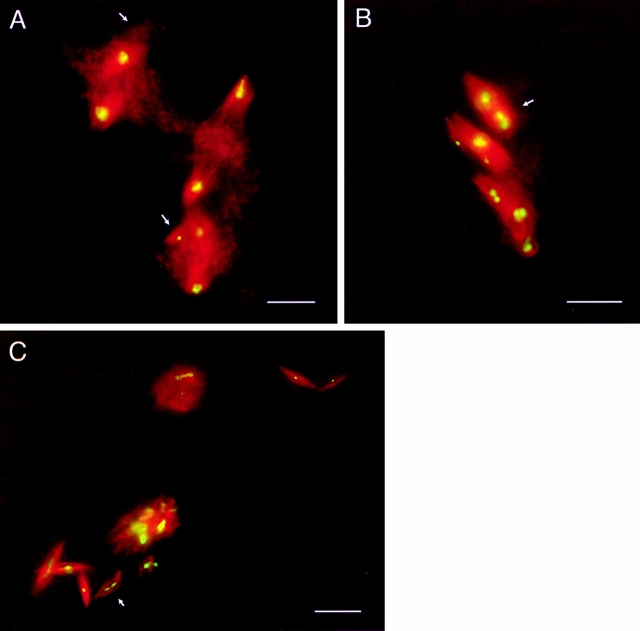
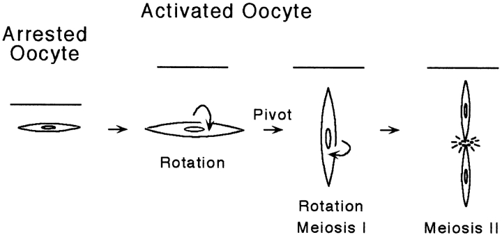
Mature oocytes of Drosophila are arrested in metaphase of meiosis I. Upon activation by ovulation or fertilization, oocytes undergo a series of rapid changes that have not been directly visualized previously. We report here the use of the Nonclaret disjunctional (Ncd) microtubule motor protein fused to the green fluorescent protein (GFP) to monitor changes in the meiotic spindle of live oocytes after activation in vitro. Meiotic spindles of metaphase-arrested oocytes are relatively stable, however, meiotic spindles of in vitro-activated oocytes are highly dynamic: the spindles elongate, rotate around their long axis, and undergo an acute pivoting movement to reorient perpendicular to the oocyte surface. Many oocytes spontaneously complete the meiotic divisions, permitting visualization of progression from meiosis I to II. The movements of the spindle after oocyte activation provide new information about the dynamic changes in the spindle that occur upon re-entry into meiosis and completion of the meiotic divisions. Spindles in live oocytes mutant for a loss-of-function ncd allele fused to gfp were also imaged. The genesis of spindle defects in the live mutant oocytes provides new insights into the mechanism of Ncd function in the spindle during the meiotic divisions.


Figures

Figure 6
Figure 6
Meiotic divisions in an activated live ncd 2–gfp* mutant oocyte. Images from a time lapse sequence of an activated live ncd 2– gfp* mutant oocyte show, from left to right and top to bottom, the spindle consisting of closely apposed separate spindles (A and B), separation of the spindle poles (C, arrows), and the formation of a small focus of Ncd2–GFP* (C, arrowhead) that marks the position of the central spindle poles. Movement of the new spindle poles away from the rest of the spindle (D–H) caused a short multipolar spindle to form (H) due to the attachment of the microtubules to the original poles. Time in minutes and seconds is indicated on each frame. Bar, 10 μm.
Figure 7
Figure 7
Release of spindle poles in an activated live ncd 2–gfp* mutant oocyte. Time lapse images of an activated live ncd 2–gfp* mutant oocyte show, from left to right and top to bottom, three spindles that were initially separated from one another moving in different directions to become somewhat more widely separated (A–C), fragmentation of spindle fibers in the centers of the spindles (D–H), and release of the spindle poles (G and H). Fluorescent particles are present in the cytoplasm and form a hazy network in the center of the spindles (E–H). These are probably microtubule fragments bound to Ncd2–GFP*. Time in minutes and seconds is indicated on each frame. Bar, 10 μm.
Figure 1
Figure 1
Meiosis I spindle in a nonactivated live ncd–gfp* oocyte. The image was taken from a time lapse series of an ncd–gfp* oocyte that was prepared by dissection under oil. The spindle was relatively stable, showing only slight changes in position and orientation during the 16.5-min observation time. Bar, 5 μm.
Figure 2
Figure 2
Metaphase I spindles in nonactivated fixed wild-type oocytes. Fixed oocytes were stained with anti–α-tubulin antibody (red) and DAPI (green) to visualize meiotic spindles and chromosomes. The position of the chromosomes in the spindle corresponds to the dark hollow in the center of the spindle in the live ncd–gfp* oocyte (Fig. 1). The dot-like fourth chromosomes (arrows) were found either (A) associated with the condensed mass of chromosomes or (B) positioned closer to the poles in the metaphase I spindles. Bar, 5 μm.
Figure 3
Figure 3
Meiotic spindle dynamics in an activated live ncd–gfp oocyte. Images from a time lapse series showing, from top to bottom, the spindle initially positioned parallel to the surface of the oocyte (A) and elongation of the spindle (B), followed by contraction and pivoting (C) into a vertical position with respect to the oocyte surface (D). The spindle remained in this position at least 9.2 min after pivoting, rotating around its long axis. Time in minutes and seconds is shown on each image. Bar, 5 μm.
Figure 4
Figure 4
Meiosis I to meiosis II progression in an activated live ncd–gfp oocyte. Time lapse images showing, from top to bottom, the meiosis I ncd–gfp oocyte spindle (A), elongation of the meiosis I spindle (B), reassembly into two tandem meiosis II spindles, and progression into meiosis II (C and D). Time in minutes and seconds is shown on each image. The arrow (B) indicates a bright focus of Ncd–GFP where the central spindle poles form. The spindles are positioned obliquely to the oocyte cortex, permitting the meiosis I and II spindles to be completely imaged. The interior of the oocyte is to the left. Bar, 10 μm.
Figure 5
Figure 5
Meiosis II spindles in normally activated fixed wild-type eggs. Normally oviposited wild-type eggs were fixed and stained with anti–α-tubulin antibody (red) and DAPI (green) to visualize the spindles and chromosomes. The meiosis II spindles are perpendicular to the cortex and are viewed down their long axis. The bottom spindle in each image, which is closer to the surface of the embryo, is slightly delayed relative to the more internal spindle; this is probably an artifact caused by the time required for penetration of the fixative. (A) Meiosis II spindles in early (bottom) and mid- (top) anaphase. The faint central spindle pole body (arrow), which consists of foci of tubulin in a ring-like array with radiating microtubules, can be observed in the region between the two spindles. (B) Meiosis II spindles in mid- (bottom) and late (top) anaphase. The central spindle pole body appears as a diffuse array of microtubules between the two spindles. Bar, 10 μm.
Figure 8
Figure 8
Meiotic divisions in normally activated fixed ncd 2–gfp* mutant eggs. Normally oviposited mutant ncd 2–gfp* eggs were fixed and stained with anti–α-tubulin antibody (red) and DAPI (green) to visualize the spindle microtubules and meiotic chromosomes. The cortex of the egg is nearer to the bottom of each of the images. (A) Late anaphase I half bivalents are segregating on separate spindles. The tiny fourth chromosomes (arrows) are associated with spindle spurs or small spindles. A hazy mass of tubulin-positive material is present in the central region of each spindle and is interpreted to be fragmented microtubules. The spindles at the top and bottom are bent or skewed, and the spindle in the middle is positioned obliquely with respect to the cortex. (B) Metaphase II chromosomes are associated with separate spindles or spindle spurs. The spindles are positioned obliquely or vertically (arrow) with respect to the cortex. The spindle indicated by the arrow is cross-sectioned and joined to a spindle spur with an associated chromosome. (C) Continued spindle-associated divisions of the maternal chromosomes after the initial two divisions. The egg contained 18 spindle-associated chromosomes or nondisjoined sister chromatids, more than the number of chromosomes expected after completion of the two meiotic divisions. Several of the chromosomes appear to be dividing. An anaphase chromosome configuration is indicated by the arrow. The spindles lack centrosomes and are present near the cortex, where the polar bodies are found in wild-type embryos. The egg was unfertilized and contained no mitotic spindles. Bars, 10 μm.
Figure 9
Figure 9
Model of spindle dynamics after oocyte activation. Meiosis I spindles in Drosophila oocytes are assembled parallel to the cortex of the oocyte (represented by the line) and remain in this position during metaphase arrest. After normal activation by ovulation, the oocyte swells. The spindle rotates around its long axis and pivots into a vertical position with respect to the cortex, followed by further rotations. Completion of meiosis I occurs with the spindle perpendicular to the cortex and is immediately followed by reorganization of the meiosis I spindle into two tandem meiosis II spindles and progression into meiosis II.

References

    1. Chalfie M, Tu Y, Euskirchen G, Ward WW, Prasher DC. Green fluorescent protein as a marker for gene expression. Science (Wash DC) 1994;263:802–805. - PubMed
    1. Chandra R, Salmon ED, Erickson HP, Lockhart A, Endow SA. Structural and functional domains of the Drosophilancd microtubule motor protein. J Biol Chem. 1993;268:9005–9013. - PubMed
    1. Doxsey SJ, Stein P, Evans L, Calarco PD, Kirschner M. Pericentrin, a highly conserved centrosome protein involved in microtubule organization. Cell. 1994;76:639–650. - PubMed
    1. Endow SA, Komma DJ. Centrosome and spindle function of the DrosophilaNcd microtubule motor visualized in live embryos using NcdGFP fusion proteins. J Cell Sci. 1996;109:2429–2442. - PubMed
    1. Endow, S.A., and D.W. Piston. 1997. Methods and protocols. In GFP: Green Fluorescent Protein Strategies and Applications. M. Chalfie and S. Kain, editors. John Wiley & Sons Inc., New York. In press.

Publication types