. 1997 Jun 16;137(6):1393-401.
doi: 10.1083/jcb.137.6.1393.

Possible involvement of phosphorylation of occludin in tight junction formation

Affiliations

Possible involvement of phosphorylation of occludin in tight junction formation

A Sakakibara et al. J Cell Biol. .

Abstract

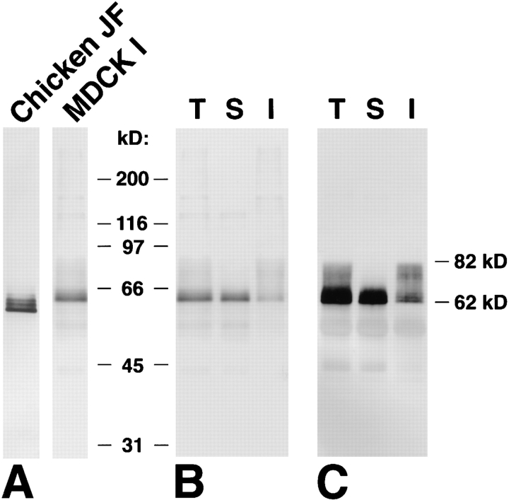
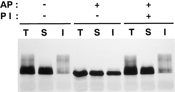
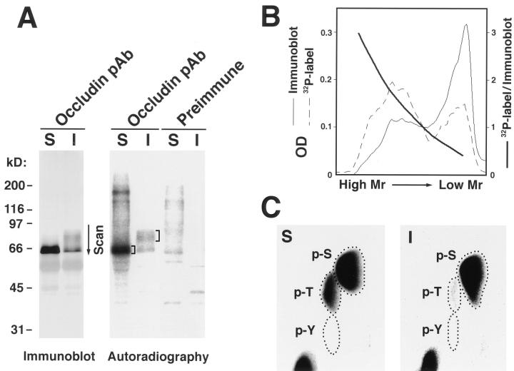
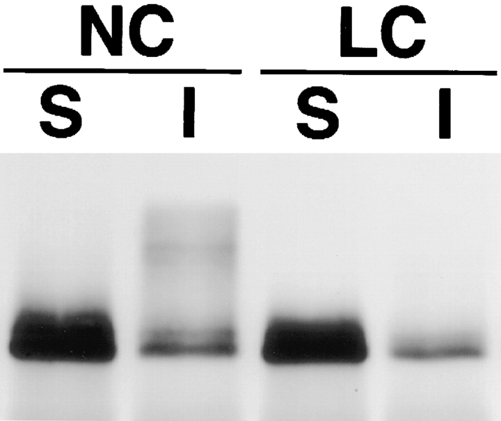
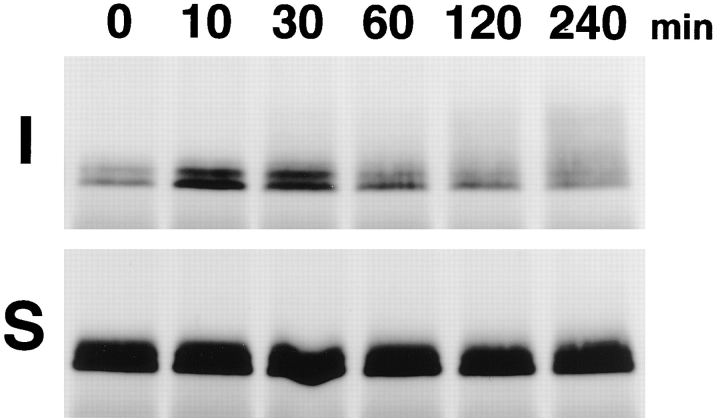
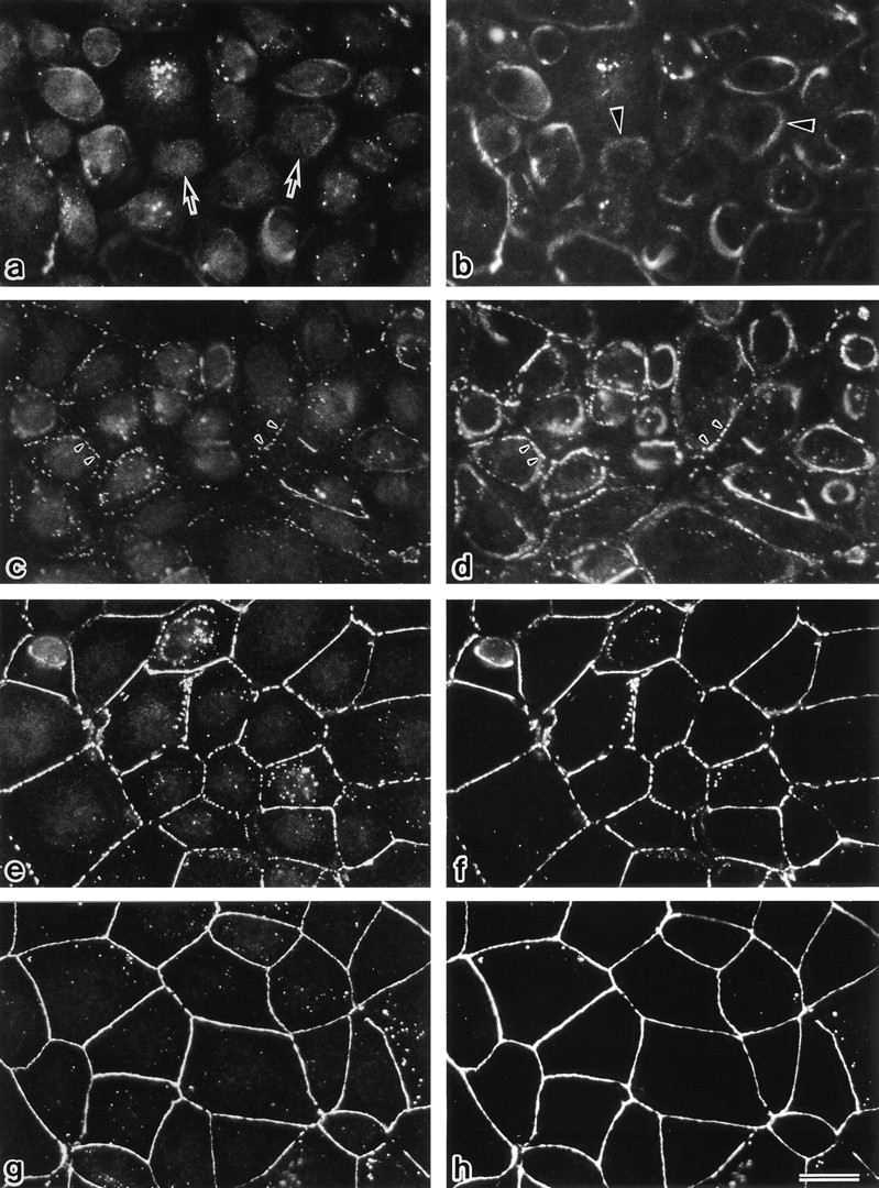
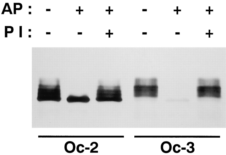
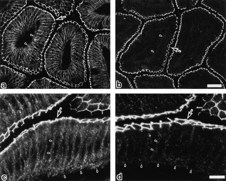
Occludin is an integral membrane protein localizing at tight junctions in epithelial and endothelial cells. Occludin from confluent culture MDCK I cells resolved as several (>10) bands between 62 and 82 kD in SDS-PAGE, of which two or three bands of the lowest Mr were predominant. Among these bands, the lower predominant bands were essentially extracted with 1% NP-40, whereas the other higher Mr bands were selectively recovered in the NP-40-insoluble fraction. Alkaline phosphatase treatment converged these bands of occludin both in NP-40-soluble and -insoluble fractions into the lowest Mr band, and phosphoamino acid analyses identified phosphoserine (and phosphothreonine weakly) in the higher Mr bands of occludin. These findings indicated that phosphorylation causes an upward shift of occludin bands and that highly phosphorylated occludin resists NP-40 extraction. When cells were grown in low Ca medium, almost all occludin was NP-40 soluble. Switching from low to normal Ca medium increased the amount of NP-40-insoluble occludin within 10 min, followed by gradual upward shift of bands. This insolubilization and the band shift correlated temporally with tight junction formation detected by immunofluorescence microscopy. Furthermore, we found that the anti-chicken occludin mAb, Oc-3, did not recognize the predominant lower Mr bands of occludin (non- or less phosphorylated form) but was specific to the higher Mr bands (phosphorylated form) on immunoblotting. Immunofluorescence microscopy revealed that this mAb mainly stained the tight junction proper of intestinal epithelial cells, whereas other anti-occludin mAbs, which can recognize the predominant lower Mr bands, labeled their basolateral membranes (and the cytoplasm) as well as tight junctions. Therefore, we conclude that non- or less phosphorylated occludin is distributed on the basolateral membranes and that highly phosphorylated occludin is selectively concentrated at tight juctions as the NP-40-insoluble form. These findings suggest that the phosphorylation of occludin is a key step in tight junction assembly.


Figures

Figure 1
Figure 1
Multiple banding pattern and detergent solubility of occludin. (A) Immunoblots of the isolated junctional fraction from the chick liver (Chicken JF) and the whole cell lysate of MDCK I cells (MDCK I) with anti–chicken occludin mAb (Oc-2) and anti–mouse occludin pAb (F4), respectively. The apparent molecular masses of chicken and dog occludin were distributed between 58 and 66 kD and 62 and 82 kD, respectively. (B) Anti-occludin pAb (F4) immunoblots of the total (T), NP-40–soluble (S), and NP-40–insoluble (I) fractions of confluent MDCK I cells (see Materials and Methods). (C) Anti-occludin mAb (MOC37) immunoblots of the anti-occludin pAb (F4 + F5) immunoprecipitates from the total (T), NP-40–soluble (S), and NP-40–insoluble (I) fractions of confluent MDCK I cells. Since the amount of occludin in each fraction was fairly small (B), both NP-40–soluble and -insoluble occludins were recovered by immunoprecipitation, electrophoresed, and immunoblotted (C). Comparison between B and C revealed that the efficiency of immunoprecipitation from NP-40–soluble fraction is almost the same as that from NP40–insoluble fraction. Higher M r bands of occludin were selectively recovered in the NP-40–insoluble fraction.
Figure 2
Figure 2
Alkaline phosphatase treatment. Anti-occludin pAb (F4 + F5) immunoprecipitates from the total (T), NP-40–soluble (S), and NP-40–insoluble (I) fractions of confluent MDCK I cells were incubated in the presence (+) or absence (−) of alkaline phosphatase (AP) and its specific inhibitor (PI) and then immunoblotted with anti-occludin mAb (MOC37). Alkaline phosphatase significantly decreased the apparent molecular masses of NP-40–soluble and -insoluble occludin bands to the level of the lowest M r band, and its inhibitor completely suppressed this effect.
Figure 3
Figure 3
Phosphoamino acid analysis of the NP-40–soluble and -insoluble occludin. (A) Anti-occludin mAb (MOC37) immunoblots (Immunoblot) and accompanying autoradiograms (Autoradiography) of anti-occludin pAb (F5) immunoprecipitates from the NP-40–soluble (S) and NP40–insoluble (I) fractions of confluent MDCK I cells metabolically labeled with [32P]orthophosphate. Control experiments were performed using preimmune serum (Preimmune). (B) Relative specific activity of occludin bands. The region marked by an arrow in the immunoblot and autoradiogram lanes of NP40–insoluble occludin was scanned by densitometry (A, Scan). Relative specific activity of each occludin band was calculated as autoradiogram density/immunoblot density. (C) The marked region in the autoradiogram lane of 32P-labeled NP-40–soluble and -insoluble occludins was excised and processed for phosphoamino acid analysis. The positions of phosphoserine (p-S), phosphothreonine (p-T), and phosphotyrosine (p-Y) were determined by autoradiography through comparison with the ninhydrin staining profiles of unlabeled phosphoamino acid standards. In NP-40–soluble occludin, both serine and threonine residues were phosphorylated (S), whereas in higher M r bands of NP-40–insoluble occludin, serine residues were predominantly phosphorylated with slight phosphorylation of threonine residues (I).
Figure 4
Figure 4
Multiple banding pattern and detergent solubility of occludin in MDCK I cells grown in normal calcium (1.8 mM; NC) or low calcium (5 μM; LC) medium for 24 h. Anti-occludin pAb (F4 + F5) immunoprecipitates from the NP-40–soluble (S) and NP-40–insoluble (I) fractions of confluent MDCK I cells were immunoblotted with anti-occludin mAb (MOC37). Under low calcium conditions, the amount of NP-40–insoluble occludin was fairly small.
Figure 5
Figure 5
Insolubilization and upward band shift of occludin in MDCK I cells after switching from low (5 μM) to normal (1.8 mM) calcium medium. 0, 10, 30, 60, 120, and 240 min after the Ca switch, the anti-occludin pAb (F4 + F5) immunoprecipitates from the NP-40–insoluble (I) or NP-40–soluble (S) fractions of confluent MDCK I cells were immunoblotted with anti-occludin mAb (MOC37). Within 10 min after switching, the amount of occludin recovered as the NP-40–insoluble portion was significantly increased, followed by a gradual increase of their apparent molecular masses.
Figure 6
Figure 6
Formation of tight junctions in MDCK I cells after a Ca switch from low (5 μM) to normal (1.8 mM) calcium medium. 0 (a and b), 10 (c and d), 30 (e and f), and 60 (g and h) min after the Ca switch, confluent MDCK I cells were fixed and immunofluorescently stained with rat anti-occludin mAb (a, c, e, and g) and mouse anti–ZO-1 mAb (b, d, f, and h). In the low calcium medium (a and b), occludin signal was detected mainly from small granular structures in the cytoplasm (arrows), and ZO-1 signal was from ring-like structures (arrowheads). Within 10 min after the Ca switch (c and d), both occludin and ZO-1 gradually began to accumulate and colocalize at cell–cell borders (arrowheads). 30–60 min after the Ca switch (e–h), both occludin and ZO-1 were colocalized in a linear fashion at junctional regions. Even 60 min after the Ca switch, the epithelial sheet was still leaky in terms of TER. Bar, 20 μm.
Figure 7
Figure 7
Two distinct anti–chicken occludin mAbs, Oc-2 and Oc-3. Chicken occludin in the isolated junctional fraction was solubilized using 1% SDS, immunoprecipitated with anti–chicken occludin pAb (F44), incubated in the presence (+) or absence (−) of alkaline phosphatase (AP) and its inhibitor (PI), and then immunoblotted with Oc-2 or -3. Oc-3 could not detect dephosphorylated occludin, whereas Oc-2 recognized both phosphorylated and dephosphorylated occludin.
Figure 8
Figure 8
Confocal immunofluorescence microscopy of frozen sections of chick intestinal epithelial cells with anti–chicken occludin mAb, Oc-2 (a and c), or Oc-3 (b and d). Oc-2 stained both the junctional complex regions (arrows) and the basolateral membrane domains (arrowheads) in linear and dotted manners, respectively. By contrast, Oc-3 mainly stained the tight junction region (arrows), showing a very weak signal only from the basolateral membranes (arrowheads). In our previous study (Furuse et al., 1993) it was emphasized that Oc-2 is specific for tight junctions without paying special attention to its staining at the basolateral membrane domains, but as shown here, the difference in the staining pattern is significant between Oc-2 and -3. Bars: (a and b) 30 μm; (c and d) 10 μm.

Similar articles

Cited by

References

    1. Anderson JM, Stevenson BR, Jesaitis LA, Goodenough DA, Mooseker MS. Characterization of ZO-1, a protein component of the tight junction from mouse liver and Madin-Darby canine kidney cells. J Cell Biol. 1988;106:1141–1149. - PMC - PubMed
    1. Ando-Akatsuka Y, Saitou M, Hirase T, Kishi M, Sakakibara A, Itoh M, Yonemura S, Furuse M, Tsukita Sh. Interspecies diversity of the occludin sequence: cDNA cloning of human, mouse, dog, and rat–kangaroo homologues. J Cell Biol. 1996;133:43–47. - PMC - PubMed
    1. Balda MS, Gonzalez-Mariscal L, Contreras RG, Macias-Silva M, Torres-Marquez ME, Garcia-Sainz JA, Cereijido M. Assembly and sealing of tight junctions: possible participation of G-proteins, phospholipase C, protein kinase C and calmodulin. J Membr Biol. 1991;122:193–202. - PubMed
    1. Balda MS, Gonzalez-Mariscal L, Matter K, Cereijido M, Anderson JM. Assembly of the tight junction: the role of diacylglycerol. J Cell Biol. 1993;123:293–302. - PMC - PubMed
    1. Balda MS, Whitney JA, Flores C, González S, Cereijido M, Matter K. Functional dissociation of paracellular permeability and transepithelial electrical resistance and disruption of the apical–basolateral intramembrane diffusion barrier by expression of a mutant tight junction membrane protein. J Cell Biol. 1996;134:1031–1049. - PMC - PubMed

Publication types