. 1997 Jun 16;185(12):2101-10.
doi: 10.1084/jem.185.12.2101.

Altered immune responses in interleukin 10 transgenic mice

Affiliations

Altered immune responses in interleukin 10 transgenic mice

A Hagenbaugh et al. J Exp Med. .

Abstract

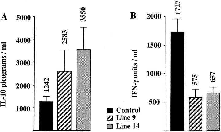
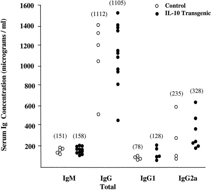
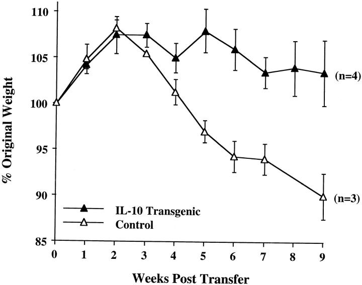
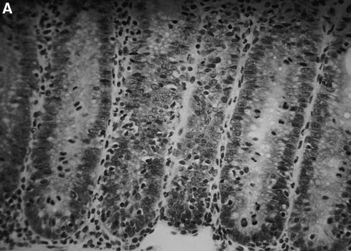
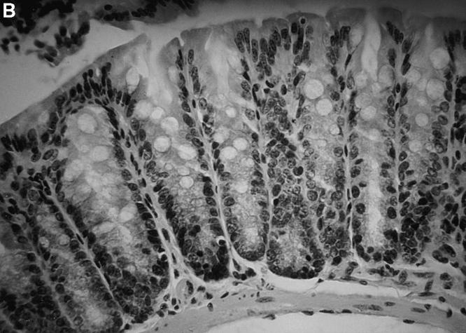
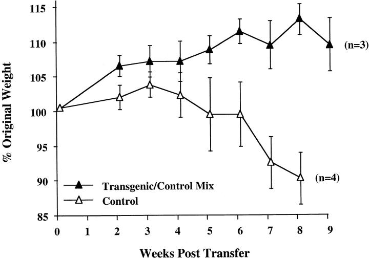
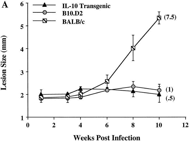
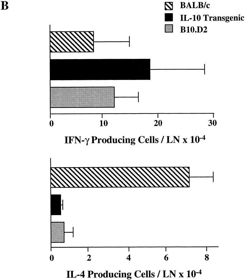
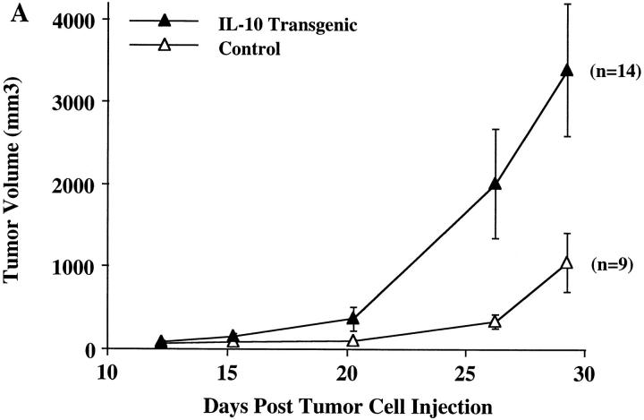
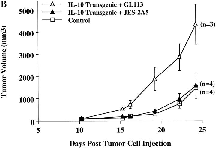
Interleukin (IL)-10 is a pleiotropic cytokine which inhibits a broad array of immune parameters including T helper cell type 1 (Th1) cytokine production, antigen presentation, and antigen-specific T cell proliferation. To understand the consequences of altered expression of IL-10 in immune models of autoimmune disease, the response to infectious agents, and the response to tumors, we developed transgenic mice expressing IL-10 under the control of the IL-2 promoter. Upon in vitro stimulation, spleen cells from unimmunized transgenic mice secrete higher levels of IL-10 and lower amounts of IFN-gamma than do controls, although no gross abnormalities were detected in lymphocyte populations or serum Ig levels. Transfer of normally pathogenic CD4(+) CD45RBhigh splenic T cells from IL-10 transgenic mice did not cause colitis in recipient severe combined immunodeficiency mice. Furthermore, co-transfer of these transgenic cells with CD4(+) CD45RBhigh T cells from control mice prevented disease. Transgenic mice retained their resistance to Leishmania major infection, indicating that their cell-mediated immune responses were not globally suppressed. Lastly, in comparison to controls, IL-10 transgenic mice were unable to limit the growth of immunogenic tumors. Administration of blocking IL-10 mAbs restored in vivo antitumor responses in the transgenic mice. These results demonstrate that a single alteration in the T cell cytokine profile can lead to dramatic changes in immune responses in a manner that is stimulus dependent. These mice will be useful in defining differences in inflammatory conditions and cellular immunity mediated by IL-10.


Figures

Figure 1
Figure 1
Altered cytokine secretion in IL-10 transgenic mice. Spleen cells from two lines of IL-10 transgenic mice and control littermates were isolated and stimulated on anti-CD3 coated plates. Supernatants were tested for the presence of IL-10 (A) and IFN-γ (B) by ELISA. Results are represented as the mean ± SE, with the mean indicated above the error bar. 3–5 mice were tested in each group.
Figure 2
Figure 2
Ig levels are not altered in IL-10 transgenic mice. Serum samples from line 9 IL-10 transgenic (filled circles) and control littermate (open circles) mice were assayed for the indicated Ig isotypes by ELISA. The group of mice analyzed for IgM and total IgG was separate from the group used for IgG1 and IgG2a analysis. The average concentration in micrograms per milliliter for each isotype is indicated in parentheses. Each symbol represents data from one individual mouse.
Figure 3
Figure 3
CD4+ CD45RBhigh cells from IL-10 transgenic mice fail to induce colitis in SCID recipients. 4–5 × 10⁵ FACS® sorted CD4+ CD45RBhigh cells from IL-10 transgenic or control littermate mice were injected into SCID mice. Weight gain/loss was scored weekly for 9 wk. Values are shown as a percentage of original weight at day 0, the time of cell injection. Number of mice in each group is indicated in parentheses. Regression analysis of the two curves shown indicated the data to be highly significant, P = 0.0012. (Similar results were obtained using Rag-2−/− mice as T cell recipients.)
Figure 4
Figure 4
Lack of intestinal inflammation in SCID recipients of CD4+ CD45RBhigh T cells from IL-10 transgenic mice. Tissues from the distal colon were stained with hematoxylin and eosin and scored as described in Materials and Methods. (A) Section of distal colon from a SCID recipient injected with control CD4+ CD45RBhigh T cells. Note the loss of goblet cells and disorganization of the epithelial cells. (B) Section of distal colon from a SCID recipient injected with IL-10 transgenic, CD4+ CD45RBhigh T cells. Original magnification, ×78 for both sections.
Figure 4
Figure 4
Lack of intestinal inflammation in SCID recipients of CD4+ CD45RBhigh T cells from IL-10 transgenic mice. Tissues from the distal colon were stained with hematoxylin and eosin and scored as described in Materials and Methods. (A) Section of distal colon from a SCID recipient injected with control CD4+ CD45RBhigh T cells. Note the loss of goblet cells and disorganization of the epithelial cells. (B) Section of distal colon from a SCID recipient injected with IL-10 transgenic, CD4+ CD45RBhigh T cells. Original magnification, ×78 for both sections.
Figure 5
Figure 5
Co-transfer of CD4+ CD45RBhigh T cells from IL-10 transgenic mice prevents colitis pathogenesis. 2 × 10⁵ CD4+ CD45RBhigh T cells from IL-10 transgenic mice were co-injected with 4 × 10⁵ cells of the same type from control mice into SCID recipients. The recipients were monitored weekly for weight loss as described in Fig. 3.
Figure 6
Figure 6
Resistance to L. major infection in IL-10 transgenic mice. Groups of four to six IL-10 transgenic, B10.D2, and BALB/c mice were inoculated in the rear footpads with promastigotes of L. major. (A) Data represent the mean lesion size ± SE for the indicated groups of mice throughout infection. Parasite burden, representing the log10 of viable Leishmania in cultured footpad homogenates after 10 wk, is indicated in parentheses (see Materials and Methods). (B) Popliteal LN cells draining the site of infection were used in ELISPOT assays for the detection of IFN-γ and IL-4. Data represent the number of cytokine-producing cells per LN ± SE for designated mice after 10 wk of infection.
Figure 6
Figure 6
Resistance to L. major infection in IL-10 transgenic mice. Groups of four to six IL-10 transgenic, B10.D2, and BALB/c mice were inoculated in the rear footpads with promastigotes of L. major. (A) Data represent the mean lesion size ± SE for the indicated groups of mice throughout infection. Parasite burden, representing the log10 of viable Leishmania in cultured footpad homogenates after 10 wk, is indicated in parentheses (see Materials and Methods). (B) Popliteal LN cells draining the site of infection were used in ELISPOT assays for the detection of IFN-γ and IL-4. Data represent the number of cytokine-producing cells per LN ± SE for designated mice after 10 wk of infection.
Figure 7
Figure 7
Uncontrolled tumor growth in IL-10 transgenic mice. (A) Groups of 9–14 IL-10 transgenic and control littermate mice were injected subcutaneously with 3LL carcinoma cells. Tumor growth was recorded on a daily basis using metric calipers. Data are shown as the mean tumor volume for each group of mice ± SE at each indicated time point. This is representative of one of three experiments, which all yielded similar results. (B) 1.5 mg of IL-10 neutralizing antibody, JES-2A5, or control isotype antibody, GL113-5E7, were administered intraperitoneally at 24 h before and 4 d after 3LL cell injection to IL-10 transgenic mice. Control littermates were injected with 3LL cells as a negative control for tumor growth. Groups of three to four mice were analyzed. Data is shown as mean tumor volume ± SE.
Figure 7
Figure 7
Uncontrolled tumor growth in IL-10 transgenic mice. (A) Groups of 9–14 IL-10 transgenic and control littermate mice were injected subcutaneously with 3LL carcinoma cells. Tumor growth was recorded on a daily basis using metric calipers. Data are shown as the mean tumor volume for each group of mice ± SE at each indicated time point. This is representative of one of three experiments, which all yielded similar results. (B) 1.5 mg of IL-10 neutralizing antibody, JES-2A5, or control isotype antibody, GL113-5E7, were administered intraperitoneally at 24 h before and 4 d after 3LL cell injection to IL-10 transgenic mice. Control littermates were injected with 3LL cells as a negative control for tumor growth. Groups of three to four mice were analyzed. Data is shown as mean tumor volume ± SE.

References

    1. Mosmann TR, Schumacher JH, Street NF, Budd R, O'Garra A, Fong TA, Bond MW, Moore KW, Sher A, Fiorentino DF. Diversity of cytokine synthesis and function of mouse CD4+T cells. Immunol Rev. 1991;123:209–229. - PubMed
    1. Fiorentino DF, Bond MW, Mosmann TR. Two types of mouse T helper cell. IV. Th2 clones secrete a factor that inhibits cytokine production by Th1 clones. J Exp Med. 1989;170:2081–2095. - PMC - PubMed
    1. Ho AS, Moore KW. Interleukin-10 and its receptor. Ther Immunol. 1994;1:173–185. - PubMed
    1. Hsieh CS, Heimberger AB, Gold JS, O'Garra A, Murphy KM. Differential regulation of T helper phenotype development by interleukins 4 and 10 in an αβ T-cellreceptor transgenic system. Proc Natl Acad Sci USA. 1992;89:6065–6069. - PMC - PubMed
    1. Ding L, Shevach EM. IL-10 inhibits mitogeninduced T cell proliferation by selectively inhibiting macrophage costimulatory function. J Immunol. 1992;148:3133–3139. - PubMed

Publication types