. 1997 Jun 16;185(12):2143-56.
doi: 10.1084/jem.185.12.2143.

Interleukin-5 expression in the lung epithelium of transgenic mice leads to pulmonary changes pathognomonic of asthma

Affiliations

Interleukin-5 expression in the lung epithelium of transgenic mice leads to pulmonary changes pathognomonic of asthma

J J Lee et al. J Exp Med. .

Abstract

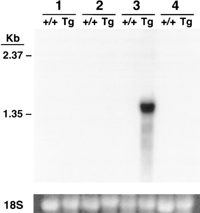
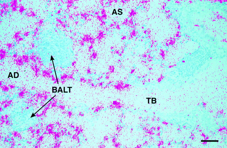
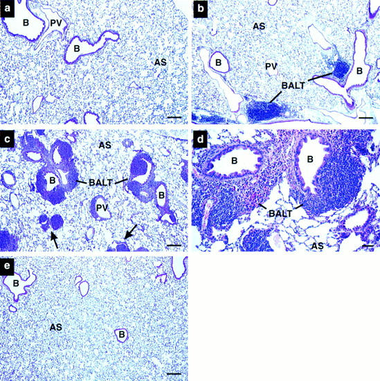
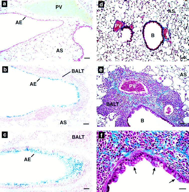
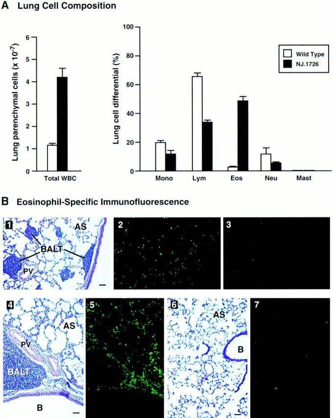
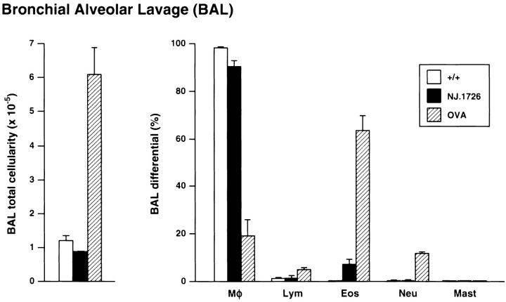
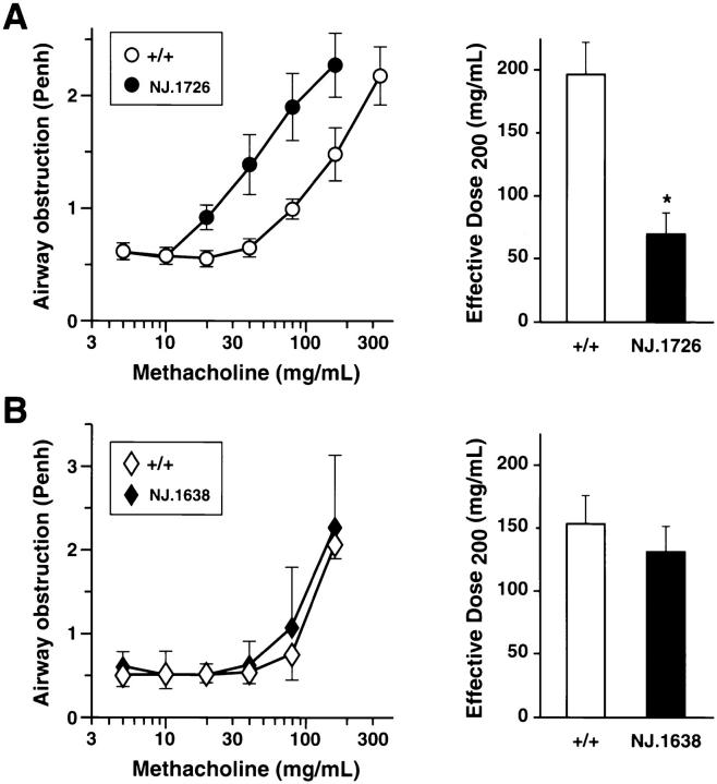
We have generated transgenic mice that constitutively express murine interleukin (IL)-5 in the lung epithelium. Airway expression of this cytokine resulted in a dramatic accumulation of peribronchial eosinophils and striking pathologic changes including the expansion of bronchus-associated lymphoid tissue (BALT), goblet cell hyperplasia, epithelial hypertrophy, and focal collagen deposition. These changes were also accompanied by eosinophil infiltration of the airway lumen. In addition, transgenic animals displayed airway hyperresponsiveness to methacholine in the absence of aerosolized antigen challenge. These findings demonstrate that lung-specific IL-5 expression can induce pathologic changes characteristic of asthma and may provide useful models to evaluate the efficacy of potential respiratory disease therapies or pharmaceuticals.


Figures

Figure 1
Figure 1
Lung-specific IL-5 gene expression. Northern blot of (+/+) and NJ.1726 (Tg) tissue RNA probed with a random-primed 32P-labeled IL-5 cDNA. Each lane contains 15 μg of total RNA. Lane 1, bone marrow; lane 2, liver; lane 3, lung; lane 4, spleen. A photograph of the 18S small ribosomal subunit stained with ethidium bromide is shown to verify the presence of RNA in each lane.
Figure 2
Figure 2
Localization of IL-5 transcripts to the lung epithelium of NJ.1726 mice. An IL-5 antisense RNA probe was synthesized from an IL-5 cDNA (a kind gift of T. Honjo) cloned into the plasmid vector pBluescript KS(+). No signal was detected when a sense probe was hybridized to adjacent sections (data not shown). TB, terminal bronchiole; AD, alveolar duct; AS, alveolar space; BALT, bronchus-associated lymphoid tissue. Bar, 25 μm.
Figure 3
Figure 3
Histopathologic pulmonary changes accompany lung epithelial expression of IL-5. (a) Wild-type, (b–d) NJ.1726, and (e) a transgenic mouse expressing IL-5 from T cells (line NJ.1638 [22]) were stained with hematoxylin and eosin before bright-field photomicroscopy. b and c are representative photographs of the variation in phenotype found in NJ.1726 mice. The arrows in c indicate airways in an NJ.1726 mouse nearly occluded by the expansion of peribronchial lymphoid tissue. The high magnification view of a BALT aggregate (d) shows in greater detail the additional histopathologies associated with these regions. e demonstrates that although T cell–specific expression of IL-5 elevates serum levels to 400–800 pg/ml, no pulmonary changes occurred, and thus the pathologies occurring in NJ.1726 mice are the result of lung-specific IL-5 expression. B, bronchiole; PV, pulmonary blood vessel; AS, alveolar space; BALT, bronchus-associated lymphoid tissue. Bars: (a–c, e) 200 μm; (d) 50 μm.
Figure 4
Figure 4
Goblet cell hyperplasia, epithial hypertrophy, and collagen deposition are induced by airway expression of IL-5. (a–c) Alcian blue (pH 2.5) staining of wild-type (a) and NJ.1726 lung sections showing a large bronchus (b) and a smaller more distal bronchiole (c). Intensely blue staining areas are glycoprotein (mucin)-containing goblet cells. (d–e) Masson's trichrome staining of paraffin sections derived from wild-type (d) and NJ.1726 (e and f) lungs. The darkly blue staining extracellular material is collagen. The arrows in f indicate hypertrophy in the bronchial epithelium. AE, airway epithelium; B, bronchiole; PV, pulmonary blood vessel; AS, alveolar space; BALT, bronchus-associated lymphoid tissue. Bars: (a–e) 50 μm; (f) 25 μm..
Figure 5
Figure 5
Cell differentials and eosinophil-specific immunofluorescence demonstrate a dramatic peribronchial infiltrate. (A) Collagenase-digested perfused lungs from NJ.1726 mice (n = 4) and transgenic negative littermates (n = 3) were dispersed to single cell suspensions prior to the assessment of lung parenchymal cellularity. The total cell counts of lung parenchymal leukocytes are shown as the histograms on the left. The relative numbers of different types of leukocytes (lung cell differentials) were determined from Wright's stained cytocentrifuge preparations and are shown as the histograms on the right. These data are expressed as the means (±SD) of the percentage of each cell type derived from differentials based on 200 cells. Mono, monocytes and macrophages; Lym, lymphocytes; Eos, eosinophils; Neu, neutrophils; Mast, mast cells. (B) Parenchymal eosinophils were localized within the lung by immunofluorescence using an anti-mMBP polyclonal rabbit antiserum. Serial lung sections (4 μm) were stained with H/E (1, 4, and 6), anti-mMBP serum (2, 5, and 7), and prebleed serum (3) to visualize lung histology and eosinophil infiltration. These photographs represent serial tissue sections from a lung of an NJ.1726 mouse (1–5) or a (+/+) control animal (6 and 7). B, bronchiole; PV, pulmonary blood vessel; AS, alveolar space; BALT, bronchus-associated lymphoid tissue. Bars: 50 μm.
Figure 6
Figure 6
Infiltration of the airway lumen by eosinophils did not occur as a consequence of the pulmonary changes in NJ.1726 mice. BAL cells derived from the lungs of 7-mo-old wild-type (+/+) (n = 6), NJ.1726 (n = 3), and, for comparison, control (+/+) animals sensitized (i.p.) and challenged with aerosolized OVA (BAL cells assessed 2 d after aerosol challenge) are displayed as histograms showing total BAL cellularity (left) and the relative numbers of different types of leukocytes (BAL differentials) determined from Wright's stained cytocentrifuge preparations (right). The fractional compositions of each cell type are expressed as percentages derived from differentials of 200 cells. Data represent mean values ±SD. , macrophages; Lym, lymphocytes; Eos, eosinophils; Neu, neutrophils; Mast, mast cells.
Figure 7
Figure 7
AHR in the absence of antigen sensitization and challenge occurs only in lung-specific IL-5–expressing transgenic mice. Airway responsiveness of NJ.1726 mice (A) and transgenic mice expressing IL-5 from a T cell–specific promoter (B) was measured immediately after exposure to aerosolized methacholine. In each case, measurements were made relative to age matched (3–5 mo) transgenic negative littermates. Mice were assessed on two occasions and the average for each animal was obtained. Values reported are group means ±SD (n = 11). These data were used to derive the effective dose200 ED200 levels (i.e., 50% maximal response) shown in the histograms to the left. The ED200 for NJ.1726 transgenic mouse group was significantly different from the control wildtype mice (*P ⩽0.01).

References

    1. O'Byrne PM. Allergen-induced airway hyperresponsiveness. J Allergy Clin Immunol. 1988;81:119–127. - PubMed
    1. Empey DW, Laitinen LA, Jacobs L, Gold WM, Nadel JA. Mechanisms of bronchial hyperreactivity in normal subjects after upper respiratory tract infection. Am Rev Resp Dis. 1976;113:131–139. - PubMed
    1. McBride DE, Koenig JQ, Luchtel DL, Williams PV, Henderson W., Jr Inflammatory effects of ozone in the upper airways of subjects with asthma. Am J Resp Crit Care Med. 1994;149:1192–1197. - PubMed
    1. Krug N, Madden J, Redington AE, Lackie P, Djukanovic R, Schauer U, Holgate ST, Frew AJ, Howarth PH. T-cell cytokine profile evaluated at the single cell level in BAL and blood in allergic asthma. Am J Resp Cell Mol Biol. 1996;14:319–326. - PubMed
    1. Henderson W., Jr Role of leukotrienes in asthma. Ann Allergy. 1994;72:272–278. - PubMed

Publication types