. 1997 Jul 1;17(13):4994-5003.
doi: 10.1523/JNEUROSCI.17-13-04994.1997.

Cholecystokinin increases GABA release by inhibiting a resting K+ conductance in hippocampal interneurons

Affiliations

Cholecystokinin increases GABA release by inhibiting a resting K+ conductance in hippocampal interneurons

K K Miller et al. J Neurosci. .

Abstract

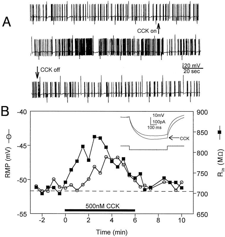
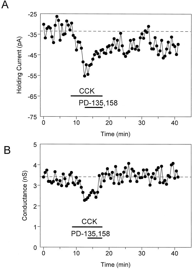
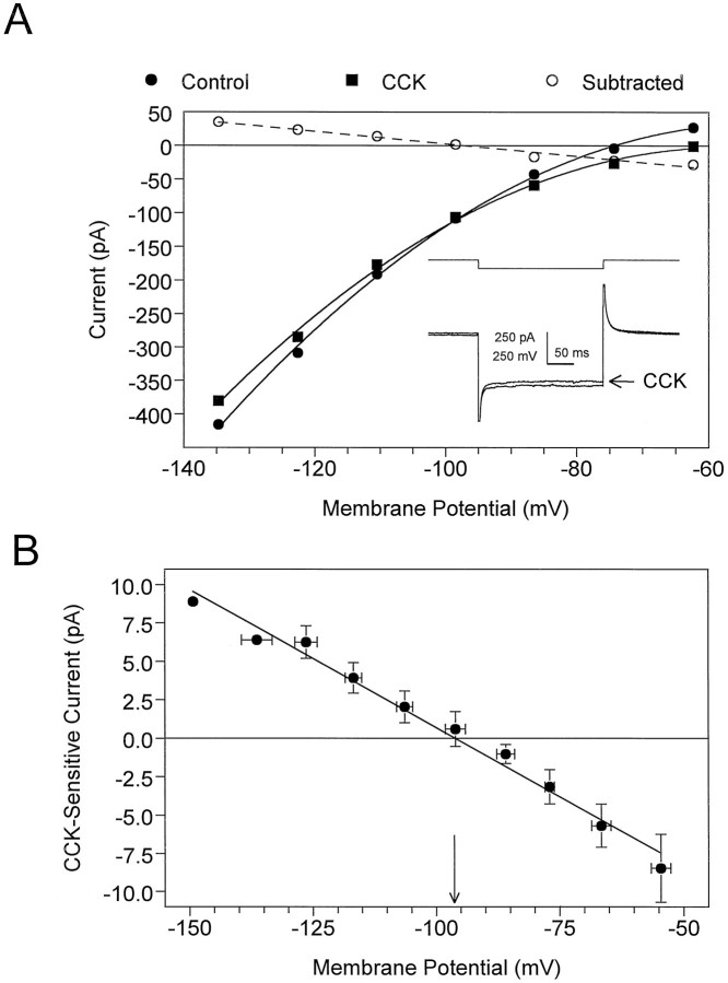
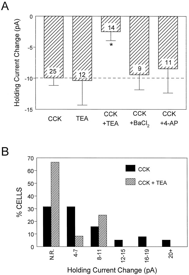
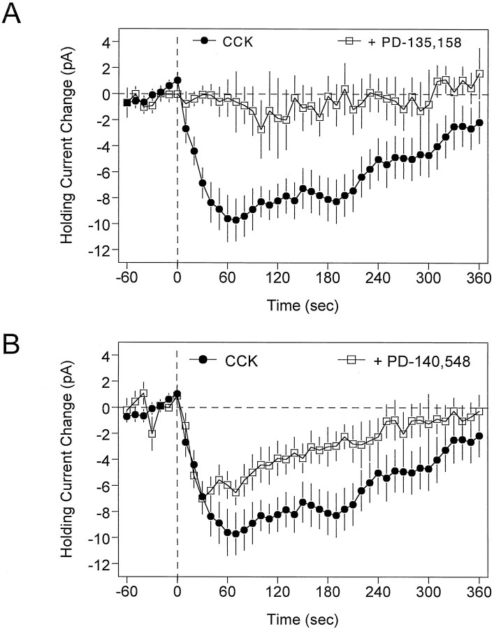
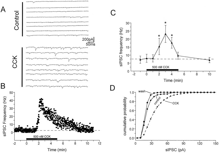
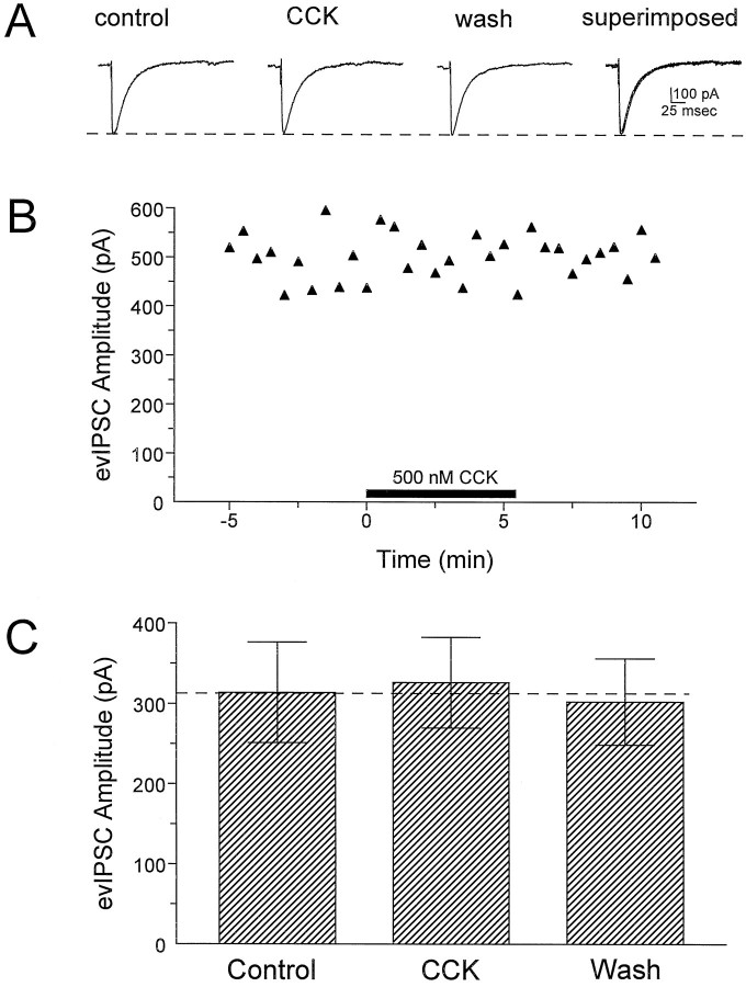
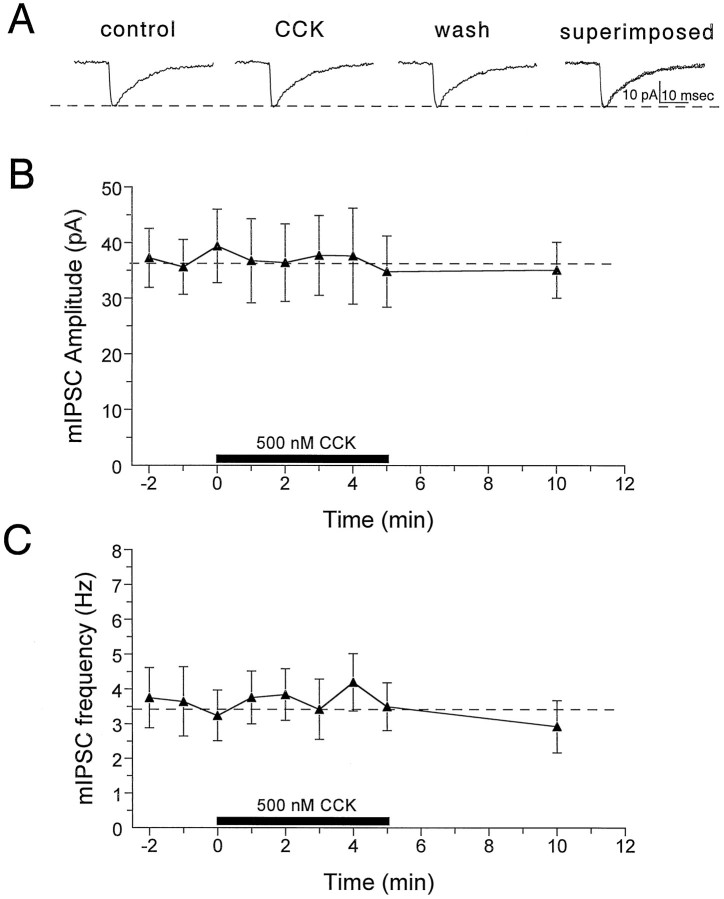
Cholecystokinin (CCK) is found co-localized with the inhibitory neurotransmitter GABA in interneurons of the hippocampus. Also, CCK receptors are found in abundance in this brain region. The possibility that CCK alters interneuron activity was examined using whole-cell current- and voltage-clamp recordings from visualized interneurons in the stratum radiatum of area CA1 in rat hippocampal slices. The effect of CCK on GABA-mediated IPSCs was also determined in pyramidal neurons. The sulfated octapeptide CCK-8S increased action potential frequency or generated inward currents in the majority of interneurons. These effects of CCK persisted in the presence of tetrodotoxin and cadmium, suggesting that they were direct. Current-voltage plots revealed that CCK-8S inhibited a conductance that was linear across command potentials and reversed near the equilibrium potential for K+ ions. The K+ channel blocker tetraethylammonium (10 mM) generated inward currents similar to those initiated by CCK, and it occluded the effect of the peptide. BaCl2 (1 mM) and 4-aminopyridine (2 mM) did not alter the effect of CCK. The CCKB receptor antagonist PD-135,158 completely blocked the inward currents generated by CCK-8S. CCK also resulted in an increase in spontaneous action potential-dependent IPSC frequency, but no changes in action potential-independent miniature IPSCs or evoked IPSCs in pyramidal neurons. These results provide evidence that CCK can depolarize hippocampal interneurons through the inhibition of a resting K+ conductance, leading to increased tonic inhibition of pyramidal neurons. This action of CCK may contribute to its anticonvulsant properties, as observed in limbic seizure models.


Figures

Fig. 1.
Fig. 1.
Whole-cell current-clamp recordings of the excitatory effects of CCK-8S (500 nm) on interneurons in the stratum radiatum of area CA1. A, Continuous chart record of the effect of CCK-8S on the action potential discharge rate in a spontaneously firing neuron. CCK-8S was bath-applied continuously beginning at the upward arrow and ending at thedownward arrow. The action potential amplitude is truncated by the slow frequency response of the chart recorder. The larger upward and downward deflections are membrane responses to depolarizing and hyperpolarizing current injection, respectively. The resting membrane potential of this cell was −54mV. B, Effects of CCK-8S (horizontal bar) on resting membrane potential (RMP, ○) and input resistance (Rin, ▪, inset) in a different stratum radiatum interneuron. Note that Rinbegins to increase just before the CCK-induced membrane depolarization. Also note that the effect of CCK-8S on firing rate (A), RMP, and Rin(B) is diminished in the continued presence of the peptide.
Fig. 2.
Fig. 2.
Effect of CCK-8S (500 nm) on holding current and whole-cell conductance in a CA1 stratum radiatum interneuron voltage-clamped at −55 mV. A, Time course illustrating the inward change in holding current with CCK-8S and the apparent reversal by the CCKB antagonist PD-135,158 (500 nm). B, Same cell as in A. Effect of CCK-8S on whole-cell steady-state membrane conductance in response to a brief hyperpolarizing voltage step (−10 mV, 300 msec). Note that the inward current caused by CCK-8S was temporally related to the decrease in whole-cell conductance. The delay in response onset was partly attributable to a 1–1.5 min lag time in the bath superfusion system.
Fig. 3.
Fig. 3.
Current–voltage (I–V) relationship obtained from stratum radiatum interneurons indicates that CCK-8S (500 nm) inhibits a voltage-independent conductance.A, The neuron was voltage-clamped at −55 mV, and the membrane was stepped from −135 mV (inset traces) to approximately −62 mV using 250 msec voltage steps. The dashed line represents a linear regression fit (r2 = 0.98) to the data obtained when the I–V relationship observed during CCK-8S superfusion was subtracted (○) from that observed before adding CCK-8S. This current is subsequently referred to as the CCK-sensitive current. TheErev for the CCK-sensitive current in this cell was −97.3 mV. Inset, Control and CCK-8S (arrow) current responses obtained at the largest voltage step. Note the decrease in the slope conductance of the I–V curve and the reduced amplitude of the current response (inset) in the presence of CCK-8S. B, Average CCK-sensitive current from four neurons. The calculatedErev from this curve was −96.1 mV (arrow), which is similar to the calculated equilibrium potential for K+ ions (−97 mV) when [K+]out = 3.0 mm.
Fig. 4.
Fig. 4.
Effect of extracellular potassium channel blockers on the CCK-induced change in holding current in stratum radiatum interneurons. A, The average ± SEM effect of CCK-8S alone and during application of the K+channel blockers TEA (10 mm), BaCl2 (1 mm), and 4-AP (2 mm) is shown. The effect of CCK-8S was significantly inhibited only by TEA (*p< 0.001). Note that TEA alone also induced an inward change in holding current that was similar in amplitude to that of CCK-8S. Thenumber superimposed on each barrepresents the number of interneurons tested in each condition.B, Frequency histogram (using the data shown inA) demonstrating that TEA decreased not only the percentage of cells responding to CCK-8S but also the magnitude of the response of those cells that did respond. N.R., No response, defined as cells exhibiting a change in holding current of ≤3 pA.
Fig. 5.
Fig. 5.
Effect of CCK receptor antagonists on CCK-induced changes in holding currents. In these experiments, the antagonist was bath-applied for 10 min, and then CCK-8S (500 nm) was bath-applied for at least 6 min beginning at time 0 (vertical dashed line). The curves represent the time course of CCK-8S effects in the absence (•) or presence (□) of the antagonist, averaged (±SEM) across at least 11 cells in each group.A, The CCKB antagonist PD-135,158 (500 nm) caused a near complete block of the effects of CCK-8S (p < 0.001). B, The CCKA antagonist PD-140,548 (200 nm) only partially antagonized the effect of CCK-8S on interneuron holding current (p < 0.05). Note the decreased effect of CCK-8S over time in the control condition, and that the response had nearly returned to baseline by 6 min of CCK-8S application.
Fig. 6.
Fig. 6.
Effects of CCK-8S on spontaneous action potential-dependent IPSCs measured in CA1 pyramidal neurons.A, Consecutive digitized current traces before (Control) and during CCK-8S (500 nm) superfusion (CCK). Whole-cell recordings of sIPSCs were performed using CsCl-filled pipettes and ionotropic glutamate receptor antagonists (Lupica, 1995). Holding potential = −80 mV. B, Time course of CCK-8S effect on sIPSC frequency recorded from the same cell represented in A. CCK-8S was bath-applied for 5 min, beginning at time 0 (solid horizontal bar). C, Time course of the effect of CCK-8S on sIPSC frequency averaged (±SEM) across six pyramidal neurons. Each point represents the average sIPSC frequency during individual 1 min periods. The time of CCK-8S application is indicated by the solid horizontal bar. In each cell tested the sIPSP frequency was significantly increased by CCK-8S (*p < 0.01 compared with control, repeated measures ANOVA). D, Cumulative probability distribution of sIPSC amplitudes, derived from the same neuron described inA and B, demonstrating a significant increase in the average sIPSC amplitude (p< 0.001, K–S test). In this particular cell the average sIPSC amplitude increased from 25.8 pA (n = 156 events) during the control period to 39.7 pA (n = 1908 events) during the third minute of CCK-8S application. A total of four of six cells showed significant sIPSC amplitude increases. Note the diminished response in the continued presence of CCK-8S (B, C).
Fig. 7.
Fig. 7.
Effects of CCK-8S on evoked IPSCs (evIPSC) in CA1 pyramidal neurons. These evoked responses were recorded simultaneously with sIPSCs using CsCl-filled electrodes and glutamate receptor antagonists (Lupica, 1995).A, Digitized averages of at least five individual responses recorded during the indicated period throughout the representative experiment shown in B. The response labeled CCK represents the average of five consecutive responses beginning at 1.5 min into the CCK-8S application. Thedashed line indicates the amplitude of the control response. B, Plot of peak evoked IPSC amplitude for a single CA1 pyramidal neuron. The period of CCK-8S application is indicated by the solid horizontal bar. C, Mean ± SEM effect of CCK-8S (500 nm) on evoked IPSCs for all cells (n = 8). The effect of CCK-8S was determined as described for the response in B. Note that CCK-8S had no effect on evoked IPSC amplitudes despite the fact that it significantly increased the frequency and amplitude of sIPSCs recorded from these same neurons (Fig. 6).
Fig. 8.
Fig. 8.
Effects of CCK-8S on mIPSCs recorded from CA1 pyramidal neurons in the presence of TTX (1 μm).A, mIPSCs averaged during 1 min periods before CCK-8S application (control, n = 90), 2 min after CCK-8S application had begun (CCK,n = 85), and 5 min after drug application had been terminated (wash, n = 88). Thedashed horizontal line represents the amplitude of the control response. Note that CCK-8S did not alter the amplitude or the kinetics of the mIPSC responses. B, Time course of the effect of CCK-8S on mIPSC amplitude averaged (±SEM) across seven pyramidal neurons. Each point represents the average mIPSC amplitude calculated during individual 1 min periods, and the time of CCK-8S application is indicated by the solid horizontal bar. The mIPSC amplitude was not significantly altered by CCK-8S in any cell (p > 0.01, K–S test).C, Time course of the effect of CCK-8S on the frequency of mIPSCs for the same group of cells described in B. In each mIPSC experiment, TTX was applied to the slice for at least 15 min before recording control events, and the efficacy of the TTX blockade of Na+ channels was assessed by monitoring the disappearance of evoked IPSCs.

Similar articles

Cited by

References

    1. Bergles DE, Doze VA, Madison DV, Smith SJ. Excitatory action of norepinephrine on multiple classes of hippocampal CA1 interneurons. J Neurosci. 1996;16:572–585. - PMC - PubMed
    1. Boden PR, Hill RG. Effects of cholecystokinin and pentagastrin on rat hippocampal neurons maintained in vitro. Neuropeptides. 1988;12:95–103. - PubMed
    1. Bohme G, Stutzmann J, Blanchard J. Excitatory effects of cholecystokinin in rat hippocampus: pharmacological response compatible with central- or B-type CCK receptors. Brain Res. 1988;451:309–318. - PubMed
    1. Bohme G, Durieux C, Stutzmann J, Charpentier B, Roques BP, Blanchard J. Electrophysiological studies with new CCK analogs: correlation with binding affinity on B-type receptors. Peptides. 1989;10:407–414. - PubMed
    1. Branchereau P, Champagnat J, Denavit-Saubie M. Cholecystokinin gated currents in neurons of the rat solitary complex in vitro. J Neurophysiol. 1993;70:2584–2595. - PubMed

Publication types

MeSH terms

LinkOut - more resources