. 1997 Jul 1;17(13):5038-45.
doi: 10.1523/JNEUROSCI.17-13-05038.1997.

Tyrosine phosphorylation of nicotinic acetylcholine receptor mediates Grb2 binding

Affiliations

Tyrosine phosphorylation of nicotinic acetylcholine receptor mediates Grb2 binding

M Colledge et al. J Neurosci. .

Abstract

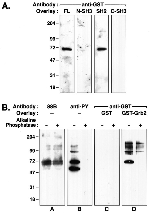
Tyrosine phosphorylation of the nicotinic acetylcholine receptor (AChR) is associated with an altered rate of receptor desensitization and also may play a role in agrin-induced receptor clustering. We have demonstrated a previously unsuspected interaction between Torpedo AChR and the adaptor protein Grb2. The binding is mediated by the Src homology 2 (SH2) domain of Grb2 and the tyrosine-phosphorylated delta subunit of the AChR. Dephosphorylation of the delta subunit abolishes Grb2 binding. A cytoplasmic domain of the delta subunit contains a binding motif (pYXNX) for the SH2 domain of Grb2. Indeed, a phosphopeptide corresponding to this region of the delta subunit binds to Grb2 SH2 fusion proteins with relatively high affinity, whereas a peptide lacking phosphorylation on tyrosine exhibits no binding. Grb2 is colocalized with the AChR on the innervated face of Torpedo electrocytes. Furthermore, Grb2 specifically copurifies with AChR solubilized from postsynaptic membranes. These data suggest a novel role for tyrosine phosphorylation of the AChR in the initiation of a Grb2-mediated signaling cascade at the postsynaptic membrane.


Figures

Fig. 1.
Fig. 1.
Grb2 binds to the δ subunit of the AChR. Torpedo postsynaptic membrane proteins (Memb) and isolated AChR subunits (AChR) were separated by SDS-PAGE and transferred to nitrocellulose membranes.A, Immunoblot with mAb 88B indicates the position of the γ and δ AChR subunits. Higher molecular weight bands are aggregates and dimers of the δ subunit and degradation products thereof. Minor lower molecular weight bands are degradation products of the γ and δ subunits. B, Tyrosine phosphorylation of the β and δ subunits of the AChR is shown by Western blotting, using anti-phosphotyrosine (anti-PY) antibody 4G10.C, Protein overlays with control GST show no binding to membrane proteins or AChR subunits. D, Overlays with GST–Grb2 show binding to a prominent band at 65 kDa, corresponding to the δ subunit of the AChR. Other bands of 90, 130, and 150 kDa in membrane samples also were observed to bind Grb2. Arrowson the left indicate the position of the β, γ, and δ AChR subunits; numbers on the rightindicate positions of molecular weight markers (in kDa).
Fig. 2.
Fig. 2.
Grb2-δ subunit binding is mediated by an SH2–phosphotyrosine interaction. A, IsolatedTorpedo AChR subunits were resolved by SDS-PAGE, transferred to nitrocellulose, and subjected to protein overlay analysis. Full-length GST–Grb2 (FL) binds to the δ subunit, as shown in Figure 1. No binding is observed with either SH3 domain fusion protein of Grb2 (N-SH3 and C-SH3). Fusion proteins containing the SH2 domain (SH2), however, bind to the δ subunit. B, Equivalent amounts ofTorpedo membranes (5 μg) treated with (right lane) and without (left lane) alkaline phosphatase were subjected to SDS-PAGE and transferred to nitrocellulose for protein overlay analysis. Panel A, Immunoblot with mAb 88B shows that approximately equal amounts of δ subunit protein were loaded for each condition.Panel B, Parallel immunoblots, using a cocktail of anti-phosphotyrosine (anti-PY) antibodies, 4G10 and PY20, indicate an absence of immunoreactivity in the phosphatase-treated membranes. Panel C, Protein overlays with control GST show no binding. Panel D, Binding of full-length GST–Grb2 to the δ subunit is abolished by phosphatase treatment. Binding to the 90 and 150 kDa bands is retained.
Fig. 3.
Fig. 3.
Tyrosine phosphorylation sites ofTorpedo AChR subunits. Alignment of tyrosine phosphorylation sites in the large cytoplasmic loop ofTorpedo β, γ, and δ subunits reveals a Grb2 SH2 consensus motif in the δ subunit, but not in the β or γ subunits. The pY+2 position is boxed and shaded. The phosphotyrosine is indicated with an arrow.
Fig. 4.
Fig. 4.
Interaction of the SH2 domain of Grb2 with a tyrosine-phosphorylated δ subunit peptide. A, Raw binding data. Resonance signal (RU) is plotted as a function of time for several concentrations of GST–Grb2 SH2 injected onto the flow cell. Binding of fusion proteins to immobilized phosphorylated (solid line) and nonphosphorylated (broken line) δ subunit peptide is shown. Concentrations of fusion proteins injected were 31.3, 62.5, 125, 250, 500, and 1000 nm. B, Determination of dissociation constant. Extrapolated steady-state binding responses (Req) are plotted versus fusion protein concentration. A dissociation constant (Kd) of 226 nm was estimated by nonlinear regression analysis.
Fig. 5.
Fig. 5.
Grb2 and AChR are colocalized in electric organ postsynaptic membranes. Frozen Narcine electric organ sections (6 μm) were labeled simultaneously for AChR with α-bungarotoxin (top, green) and Grb2 (middle, red), processed for immunofluorescence, and analyzed by confocal microscopy. Grb2 antibodies label the innervated face of the electrocyte, in precise register with the AChR (bottom, merged image).
Fig. 6.
Fig. 6.
Grb2 associates with the AChR in situ. AChRs were isolated from solubilizedTorpedo membranes by α-bungarotoxin–Sepharose, were eluted in SDS, and were resolved by SDS-PAGE. Coomassie staining of the preparation shows the four subunits of the AChR: α, ∼40 kDa; β, ∼50 kDa; γ, ∼60 kDa; and δ, ∼65 kDa. Preincubation with excess α-bungarotoxin (25 μm) prevents binding of AChR to the toxin–Sepharose (left). Immunoblotting the samples with anti-Grb2 antibodies reveals that Grb2 specifically copurifies with the AChR (right).

Similar articles

Cited by

References

    1. Blaikie P, Immanuel D, Wu J, Li N, Yajnik V, Margolis B. A region in Shc distinct from the SH2 domain can bind tyrosine-phosphorylated growth factor receptors. J Biol Chem. 1994;269:32031–32034. - PubMed
    1. Bowe MA, Deyst KA, Leszyk JD, Fallon JR. Identification and purification of an agrin receptor from Torpedo postsynaptic membranes: a heteromeric complex related to the dystroglycans. Neuron. 1994;12:1173–1180. - PubMed
    1. Campanelli JT, Roberds SL, Campbell KP, Scheller RH. A role for dystrophin-associated glycoproteins and utrophin in agrin-induced AChR clustering. Cell. 1994;77:663–674. - PubMed
    1. Chardin P, Cussac D, Maignan S, Ducruix A. The Grb2 adaptor. FEBS Lett. 1995;369:47–51. - PubMed
    1. Chavez RA, Hall ZW. Expression of fusion proteins of the nicotinic acetylcholine receptor from mammalian muscle identifies the membrane-spanning regions in the α and δ subunits. J Cell Biol. 1992;116:385–393. - PMC - PubMed

Publication types