. 1997 Jul 1;17(13):5101-7.
doi: 10.1523/JNEUROSCI.17-13-05101.1997.

Neuronal localization of presenilin-1 and association with amyloid plaques and neurofibrillary tangles in Alzheimer's disease

Affiliations

Neuronal localization of presenilin-1 and association with amyloid plaques and neurofibrillary tangles in Alzheimer's disease

J Busciglio et al. J Neurosci. .

Abstract

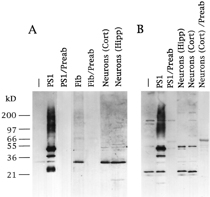
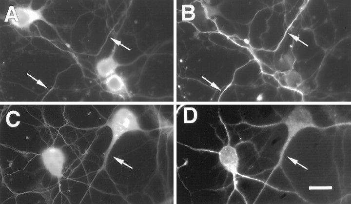
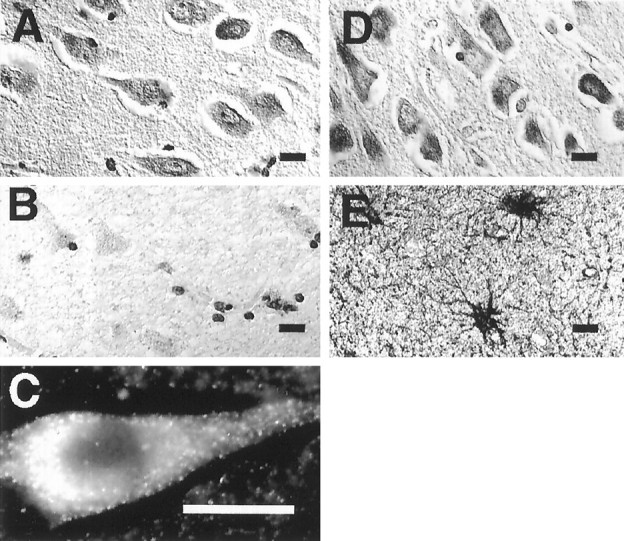
Mutations in the presenilin-1 (PS1) gene is a cause of early- onset familial Alzheimer's disease (AD). Endogenous PS1 is associated with the endoplasmic reticulum in the cell body of undifferentiated SH-SY5Y neuroblastoma cells. At early stages of neuronal differentiation in rat hippocampal culture, PS1 appears in all neuritic processes and in growth cones. In mature differentiated neurons, PS1 is concentrated in the somatodendritic compartment but is also present at lower levels in axons. A similar localization of PS1 is observed in vivo in neurons of the adult human cerebral cortex. In sporadic AD, PS1 appears in the dystrophic neurites of mature amyloid plaques and co-localizes with a subset of intraneuronal neurofibrillary tangles (NFTs). About 30% of hippocampal NFTs are labeled with a highly specific antibody to the PS1 C-terminal loop domain but not with an antibody to the PS1 N terminus. This observation is consistent with a potential association of the PS1 C-terminal fragment with NFTs, because PS1 is constitutively cleaved to N- and C-terminal fragments in neurons. These results suggest that PS1 is highly expressed and broadly distributed during early stages of neuronal differentiation, consistent with a role for PS1 in neuronal differentiation. Furthermore, the co-localization of PS1 with NFTs and plaque dystrophic neurites implicates a role for PS1 in the diverse pathological manifestations of AD.


Figures

Fig. 1.
Fig. 1.
Expression of PS1 in transfected and primary cell cultures. A, Western blot analysis of cell lysates with Ab 231 directed against the PS1 N terminus. B, Western blot analysis with Ab R28 directed against the PS1 C-terminal loop.Lanes: −, wild-type COS cells; PS1, PS1-transfected COS cells; PS1/Preab, PS1-transfected COS cells blotted with preabsorbed antibody; Fib, primary human fibroblasts; Fib/preab, fibroblasts blotted with preabsorbed antibody; Neurons (Cort), cultured human cortical neurons; Neurons (Cort)/Preab, cultured human cortical neurons blotted with preabsorbed antibody;Neurons (Hipp), cultured rat hippocampal neurons. Note the predominant 28–30 kDa N-terminal and 20–22 kDa C-terminal PS1 fragments in nontransfected cells and primary neurons.
Fig. 2.
Fig. 2.
PS1 is a resident protein of the endoplasmic reticulum in SH-SY5Y neuroblastoma cells. A, SH-SY5Y neuroblastoma cells show a reticular pattern of PS1 immunoreactivity (Ab R28) characteristic of the ER. B, C, Localization of PS1 in the endoplasmic reticulum is confirmed by double label immunofluorescence with the PS1 C-terminal loop antibody R28 (B) and anti-BiP (C). Confocal microscopy demonstrates the overlap of both antigens in an image section taken 5 μm above the substrate plane. PS1 does not co-localize with the Golgi marker JE4 in SH-SY5Y cells (data not shown). Scale bars, 10 μm.
Fig. 3.
Fig. 3.
Localization of PS1 during the initial stages of neuronal differentiation. A, Rat hippocampal neurons after 1 d in culture show PS1 immunoreactivity homogeneously distributed in the cell body and primary neuritic processes. Immunocytochemistry was performed with Aβ 231 to the PS1 N terminus. Scale bar, 10 μm. B, Examination at a higher magnification shows that PS1 is present in neuritic growth cones. Scale bar, 3 μm. C, Negative immunostaining is obtained after preabsorption of the antibody with the antigenic PS1 peptide.
Fig. 4.
Fig. 4.
Polarized distribution of PS1 after neuronal differentiation. Rat hippocampal neurons were cultured for 10 d and then double labeled for PS1 using Ab R28 to the PS1 C-terminal loop (A) and tau using Ab tau-1 (B). Arrows show co-localization of PS1 and tau in axons. Double labeling for PS1 (C) and MAP-2 (D) using Ab AP20 shows that PS1 is predominantly localized to the somatodendritic compartment (arrows) in differentiated neurons. Scale bars, 10 μm.
Fig. 5.
Fig. 5.
Localization of PS1 in the human brain.A, PS1 immunoreactivity in the perikarya of pyramidal neurons in layer CA1 of the hippocampus. Shown is immunocytochemical staining with the PS1 C-terminal loop antibody R28. B, Preabsorption of Ab R28 with the PS1 fusion protein (PS1 residues 263–407) abolishes neuronal immunoreactivity. Cellular profiles are still detected by the Nissl counterstain. C, Immunofluorescence microscopy with Ab 231 at a higher magnification shows PS1 immunoreactivity in the cytoplasm and primary dendrite of a pyramidal neuron. Adjacent sections of hippocampus labeled with PS1 Ab 231 (D) and anti-GFAP (E) demonstrate the absence of detectable PS1 in astrocytes. Sections were derived from the hippocampus of a normal 60-year-old man. Scale bar, 10 μm.
Fig. 6.
Fig. 6.
Differential labeling of NFTs by antibodies to the N- and C-terminal loop domains of PS1. Paraffin sections of hippocampus from a case of sporadic late-onset AD were immunocytochemically labeled with antibodies to phosphorylated tau and PS1. A, Labeling of an NFT with antibody PHF-1 to phosphorylated tau.B, Same region of an adjacent section immunostained with Ab R28 to the PS1 C-terminal loop labels an NFT with similar morphology. C, Section showing several NFTs (arrows) labeled by PS1 Ab R28. D, Section adjacent to the one shown in C immunostained with Ab 231 to the PS1 N terminus shows somatodendritic staining (arrow). NFTs are not labeled.
Fig. 7.
Fig. 7.
Association of PS1 with amyloid plaques and NFTs in AD. Shown are sections of hippocampus from an individual with sporadic AD. A, Double labeling with Ab 231 to PS1 (brown) and the monoclonal antibody β1 to Aβ (red) shows that PS1-positive neurons (arrow) surround amyloid plaques (arrowhead). B, Analysis of a single mature plaque by double label immunofluorescence for PS1 (green) and Aβ (dark blue). Note that PS1 appears in dystrophic neurites, which are labeledgreen for PS1 or light blue from the overlap of PS1 and Aβ. A low level of PS1 immunoreactivity appears in the amyloid core (arrowhead), which is staineddark blue for Aβ. C, Double labeling for phosphorylated tau (dark blue, Ab PHF-1) and the N-terminal epitope of PS1 (green, Ab 231). Two neurons with NFTs (arrows) are labeled for both phosphorylated tau and PS1, but the N-terminal PS1 Ab does not double label NFTs, which are labeled with Ab PHF-1. D, Double labeling for phosphorylated tau (dark blue, Ab PHF-1) and the C-terminal loop region of PS1 (green, Ab R28). Shown is an NFT that is double-labeled (arrow, light blue). An example of a neuron showing predominantly PHF-1 labeling (dark blue) is seen in the bottom right corner.

References

    1. Busciglio J, Yankner BA. Apoptosis and increased generation of reactive oxygen species in Down’s syndrome neurons in vitro. Nature. 1995;378:776–779. - PubMed
    1. Busciglio J, Gabuzda DH, Matsudaira P, Yankner BA. Generation of β-amyloid in the secretory pathway in neuronal and non-neuronal cells. Proc Natl Acad Sci USA. 1993;90:2092–2096. - PMC - PubMed
    1. Cook DG, Sung JC, Golde TE, Felsenstein KM, Wolczyk BS, Tanzi RE, Trojanowski JQ, Lee VM-Y, Doms RW. Expression and analysis of presenilin 1 in a human neuronal system: localization in cell bodies and dendrites. Proc Natl Acad Sci USA. 1996;93:9223–9226. - PMC - PubMed
    1. Cribbs DH, Chen LS, Bende SM, LaFerla FM. Widespread neuronal expression of the presenilin-1 early-onset Alzheimer’s disease gene in murine brain. Am J Pathol. 1996;148:1797–1806. - PMC - PubMed
    1. Doan A, Thinakaran G, Borchelt DR, Slunt HH, Ratovitsky T, Podlisny M, Selkoe DJ, Seeger M, Gandy SE, Price DL, Sisodia SS. Protein topology of presenilin 1. Neuron. 1996;17:1023–1030. - PubMed

Publication types