. 1997 Jul 1;17(13):5196-205.
doi: 10.1523/JNEUROSCI.17-13-05196.1997.

Mice lacking metabotropic glutamate receptor 5 show impaired learning and reduced CA1 long-term potentiation (LTP) but normal CA3 LTP

Affiliations

Mice lacking metabotropic glutamate receptor 5 show impaired learning and reduced CA1 long-term potentiation (LTP) but normal CA3 LTP

Y M Lu et al. J Neurosci. .

Abstract

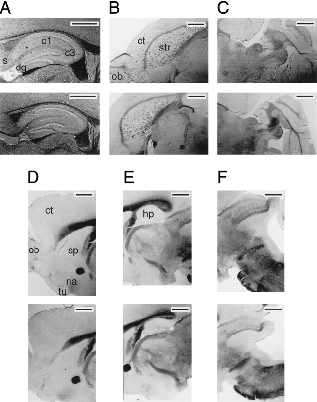
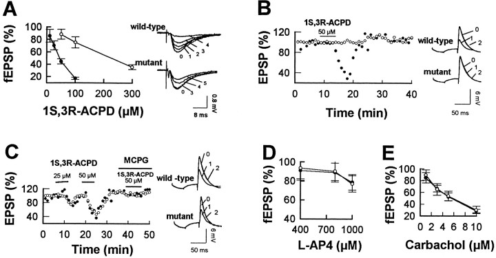
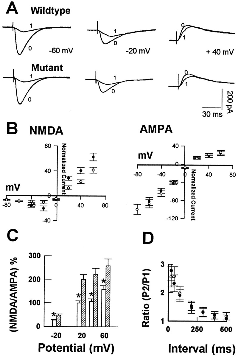
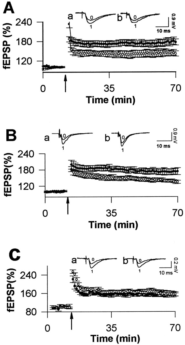
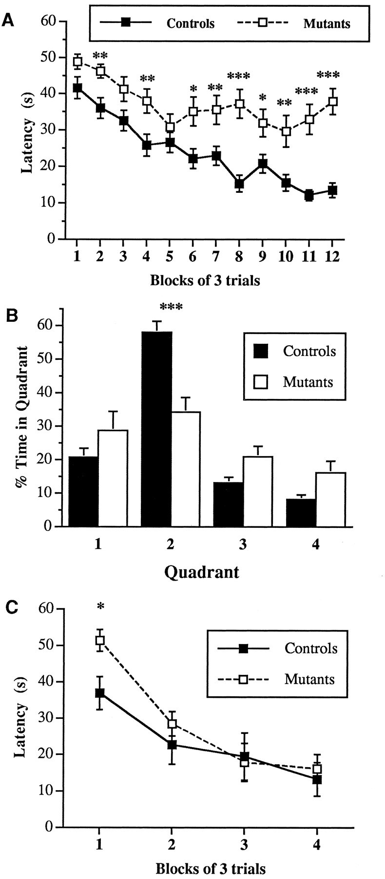
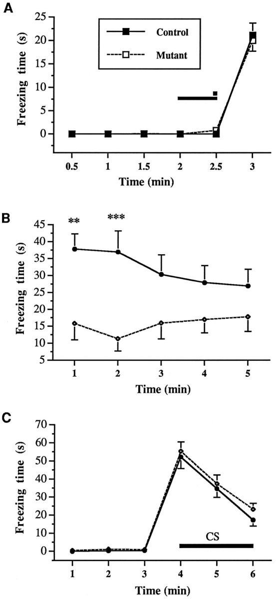
Class I metabotropic glutamate receptors (mGluRs) have been postulated to play a role in synaptic plasticity. To test the involvement of one member of this class, we have recently generated mutant mice that express no mGluR5 but normal levels of other glutamate receptors. The CNS revealed normal development of gross anatomical features. To examine synaptic functions we measured evoked field EPSPs in the hippocampal slice. Measures of presynaptic function, such as paired pulse facilitation in mutant CA1 neurons, were normal. The response of mutant CA1 neurons to low concentrations of (1S,3R)-1-amino-cyclopentane-1,3-dicarboxylic acid (ACPD) was missing, which suggests that mGluR5 may be the primary high affinity ACPD receptor in these neurons. Long-term potentiation (LTP) in mGluR5 mutants was significantly reduced in the NMDA receptor (NMDAR)-dependent pathways such as the CA1 region and dentate gyrus of the hippocampus, whereas LTP remained intact in the mossy fiber synapses on the CA3 region, an NMDAR-independent pathway. Some of the difference in CA1 LTP could lie at the level of expression, because the reduction of LTP in the mutants was no longer observed 20 min after tetanus in the presence of 2-amino-5-phosphonopentanoate. We propose that mGluR5 plays a key regulatory role in NMDAR-dependent LTP. These mutant mice were also impaired in the acquisition and use of spatial information in both the Morris water maze and contextual information in the fear-conditioning test. This is consistent with the hypothesis that LTP in the CA1 region may underlie spatial learning and memory.


Figures

Fig. 1.
Fig. 1.
Neuroanatomy in mGluR5 mice: pairwise comparisons of adult mGluR5+/− (top panels) and mGluR5−/− (bottom panels) littermates. Thirty micrometer cryostat sections at comparable levels are shown. A, Parasagittal sections showing the hippocampal formation (right, rostral;left, caudal). B, Horizontal sections through the forebrain and thalamus (2.8 mm ventral to the dorsal aspect of brain). C, Horizontal sections through the hindbrain (4.5 mm ventral to the dorsal aspect of brain). D, Parasagittal sections of the forebrain (300 μm from midline).E, Parasagittal sections showing structures of the diencephalon, hippocampus, and splenium of the corpus callosum (300 μm from midline). F, Parasagittal sections of the caudal aspect of the brain, showing regions of the superior and inferior colliculus and brainstem (450 μm from the midline). Scale bars, A, 500 μm; B–F, 1000 μm. Labeled structures represent regions previously shown to express high levels of mGluR5. c1, Hippocampal subfield CA1;c3, hippocampal subfield CA3; ct, neocortex; dg, dentate gyrus; hp, hippocampus; na, nucleus accumbens; ob, olfactory bulb; s, subiculum; sp, medial septal region (low in mGluR5 expression); str, striatum;tu, olfactory tubercle.
Fig. 2.
Fig. 2.
Decreased response to ACPD in mGluR5 mutants.A, Mean ± SEM depression of fEPSP slope induced by ACPD. Data are expressed as mean percentages of the control values in mutant mice (○, six slices, six animals) and wild-type mice (•, six slices, six animals). Traces taken from representative experiments show the effects of ACPD on evoked fEPSP. Each trace is an average of six sweeps recorded immediately before drug application (0) or after 10 min in the agonist at concentrations of 10 (1), 25 (2), 50 (3), 100 (4), and 300 (5) μm.B, Whole-cell current-clamp recording showing the time course of EPSP depression by bath application of ACPD in a single wild-type (•) or mutant (○) CA1 cell, representative of four cells. The constant current (20 pA) hyperpolarizing pulse preceding the EPSP did not give any evidence of the input resistance changes during ACPD applications. C, Representative time courses of EPSP depression in whole-cell current-clamp recordings in the medial perforant pathway of the dentate granule neurons in the presence or absence of ACPD and 1 mm MCPG in wild-type (•) and mutant (○) mice. Insets in B, C,Representative EPSP before (0), during (1), and after (2) ACPD application.D, Mean ± SEM percent fEPSP afterl-AP4 application to CA1 from control (•) and mutant (○) mice (n = 4). E, Mean fEPSP after carbachol addition to control (•) and mutant (○) mice (six slices, three animals).
Fig. 3.
Fig. 3.
Reduced NMDA component of synaptic transmission in hippocampal slices from mGluR5 mutant mice. A, The EPSC traces were recorded from CA1 neurons in whole-cell voltage-clamp mode and were averages of six successive sweeps before (0) and 20 min after (1) the addition of 10 μmCNQX. The holding membrane potentials are indicated between the traces.B, Averaged amplitudes of AMPA- and NMDA-mediated responses in mGluR5+/+ (•) and mGluR5−/− (○) mice, normalized to the 5 msec peak of AMPA EPSC at −80 mV, which was 342.6 ± 38 pA (n = 10) in control and 318.8 ± 34 pA (n = 11) in mutant mice. The AMPA component at −80 mV was taken as 100%, and all other current amplitudes were scaled and expressed as a percentage of the AMPA current. C, Data in B shown as the NMDA component of EPSCs that differed between wild-type (hatched bars) and mutants (open bars). *Significant difference (p < 0.01, t test).D, Magnitude of the paired pulse facilitation of fEPSPs in the CA1 area of the two groups (six slices from three animals for each genotype). P1, First response; P2, second response applied at the indicated intervals on thex-axis.
Fig. 4.
Fig. 4.
Reduced LTP in NMDA-dependent pathways in mGluR5 mutant mice. The mean ± SEM of the 5–95% slope of the fEPSP, normalized with respect to 10 min immediately preceding the tetanus (↑) for hippocampal slices obtained from control (•) or mutant (○) mice in area CA1 (A), the dentate gyrus medial perforant pathway (B), and the CA3 mossy fiber pathway (C). LTP in CA1 was induced by four trains of 100 Hz tetanic stimulation. LTP in the dentate gyrus was induced by four trains of tetanus in the presence of 100 μm picrotoxin. LTP in CA3 was induced by one tetanic train in the presence of 50 μm AP5. Representative traces (average of six sweeps) of fEPSP obtained immediately before (0) and 60 min after (1) the tetanus are shown for a control mouse (a) and mutant mouse (b), respectively.
Fig. 5.
Fig. 5.
Performance of mGluR5 mutants was impaired in the water maze. A, mGluR5 mutants and control mice were trained with two blocks of three trials per day (for 6 d) in the water maze. The average time to reach the hidden platform in the pool was plotted against three-trial blocks. A significant difference was found between groups. B, Percentage of time spent searching in each quadrant of the pool during the probe trail given 1 d after the last training trial. Quadrants: 2, target quadrant (southeast); 1, adjacent to the right (northeast); 3, adjacent to the left (southwest); 4, opposite (northwest). Control animals searched selectively, and significantly longer, for the platform in the training quadrant (2) than mutants.C, The average time to reach the visible platform in each three-trial block (2 d of testing) is presented. ANOVA with repeated measures did not reveal any significant differences between mutants and controls. *p < 0.05; **p < 0.01; ***p < 0.001.
Fig. 6.
Fig. 6.
Contextual fear conditioning is impaired in mGluR5 mutants. A, Duration of freezing during the training phase of fear conditioning. Mutant mGluR5 and control mice showed a comparable amount of freezing immediately after the foot shock. Thesolid line indicates the duration of the tone (CS); squares indicate the 2 sec footshock (US). B, The mice were tested for contextual conditioning 24 hr after training. Mutant mice showed significantly less freezing than controls when returned to the training chamber.C, A control tone (CS) conditioning test was carried out in a new context 2 hr after the context test. Both mutants and controls showed no freezing in a new context and comparable amounts of freezing when a tone (CS) was presented in a new context. **p < 0.01, ***p < 0.001.

Similar articles

Cited by

References

    1. Abe T, Sugihara H, Nawa H, Shigemoto R, Mizumo W, Nahanishi S. Molecular characterization of a novel metabotropic glutamate receptor, mGluR5, coupled to inositol phosphate/Ca2+ signal transduction. J Biol Chem. 1992;267:13361–13368. - PubMed
    1. Abeliovitch A, Paylor R, Chen C, Kim J, Wehner JM, Tonegawa S. PKCγ-mutant mice exhibit mild deficits in spatial and contextual learning. Cell. 1993;75:1263–1271. - PubMed
    1. Aiba A, Chen C, Herrup K, Rosenmund C, Stevens CF, Tonegawa S. Reduced hippocampal long-term potentiation and context-specific deficit in associative learning in mGluR1 mutant mice. Cell. 1994;79:365–375. - PubMed
    1. Bach ME, Hawkins RD, Osuman M, Kendel ER, Mayford M. Impairment of spatial but not contextual memory in CaMKII mutant mice with a selective loss of hippocampal LTP in the range and theta frequency. Cell. 1995;81:905–910. - PubMed
    1. Bannerman DM, Good MA, Butcher SP, Ramsay M, Morris RG. Distinct components of spatial learning revealed by prior training and NMDA receptor blockade. Nature. 1995;378:182–186. - PubMed

Publication types

MeSH terms

Substances