. 1997 Jun 24;94(13):6629-34.
doi: 10.1073/pnas.94.13.6629.

Covalent modification of the active site threonine of proteasomal beta subunits and the Escherichia coli homolog HslV by a new class of inhibitors

Affiliations

Covalent modification of the active site threonine of proteasomal beta subunits and the Escherichia coli homolog HslV by a new class of inhibitors

M Bogyo et al. Proc Natl Acad Sci U S A. .

Abstract

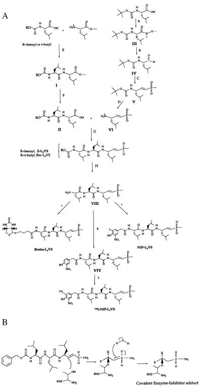
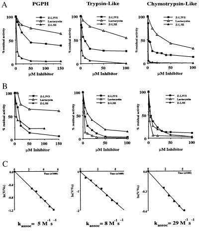
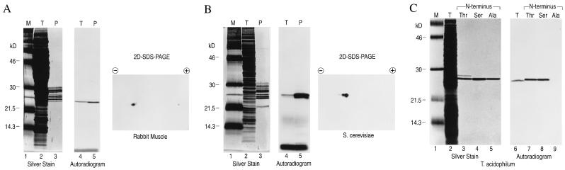
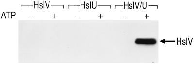
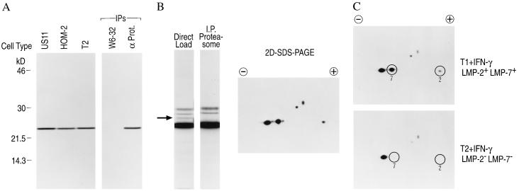
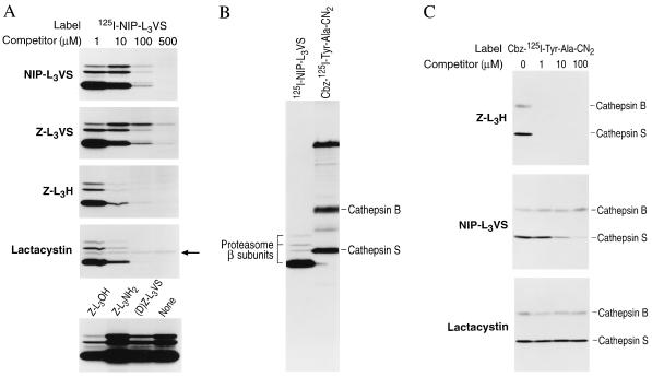
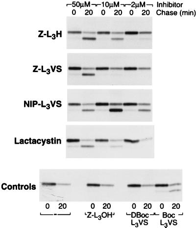
The proteasome is a multicatalytic protease complex that plays a key role in diverse cellular functions. The peptide vinyl sulfone, carboxybenzyl-leucyl-leucyl-leucine vinyl sulfone (Z-L3VS) covalently inhibits the trypsin-like, chymotrypsin-like and, unlike lactacystin, also the peptidylglutamyl peptidase activity in isolated proteasomes, and blocks their function in living cells. Although described as a class of mechanism-based inhibitors for cysteine proteases, the peptide vinyl sulfone Z-L3VS and a 125I-labeled nitrophenol derivative (125I-NIP-L3VS) covalently modify the active site threonine of the catalytic beta subunits of the proteasome. Modification of Thermoplasma proteasomes demonstrates the requirement for a hydroxyl amino acid (threonine, serine) as nucleophile at the beta subunit's NH2 terminus. 125I-NIP-L3VS covalently modifies the HslV subunit of the Escherichia coli protease complex HslV/HslU, a reaction that requires ATP, and supports a catalytic mechanism shared with that of the eukaryotic proteasome.


Figures

Figure 1
Figure 1
Synthesis (A) and mechanism of inhibition (B) of the peptide vinyl sulfones. (A) PyBOP, DIEA, CH3NHOCH3, CH2Cl2; (B) LAH, Et2O (C) (EtO)2P(O)CH2S(O)2CH3, NaH; TsOH, Et2O; (E) DCC, HOBT, DMF; (F) 20% K2CO3/MeOH; (G) PyBOP, DIEA, CH2Cl2; (H) TsOH, Et2O (I) Biotin-NP ester, DMF, DIEA; (J). 4-Hydroxy-3-iodo-5-nitrophenylacetic acid, PyBOP, DIEA, CH2Cl2; (K) 4-hydroxy-3-nitrophenylacetic acid, PyBOP, DIEA, CH2Cl2; (L) Na125I, Iodogen.
Figure 2
Figure 2
The peptide vinyl sulfones inhibit the proteasome in vitro. Purified preparations of 20S and 26S proteasomes were incubated with increasing concentrations of the three inhibitors [Z-L3H, Z-L3VS, and lactacystin either simultaneously with substrates (A)], or samples were preincubated for 1 hr (37°C) and diluted 10-fold prior to addition of substrates (B). Hydrolysis of substrates corresponding to the acidic (Cbz-LLE-βNA), basic (Boc-LRR-MCA), and hydrophobic (Suc-LLVY-MCA) activities of the proteasome were measured by fluorescence spectroscopy. Kassoc for the compound, Z-L3VS, were determined for the three peptidase activities of purified 20S proteasomes (C). V represents velocity of the reaction at the time (t), and Vo is the velocity at time 0. Kassoc = ln(V/Vo)/[I] where [I] is the concentration of inhibitor.
Figure 3
Figure 3
The peptide vinyl sulfone, 125I-NIP-L3VS, labels multiple β subunits of the proteasome in total cell extracts and in purified proteasome preparations. Total cell extracts (T) and purified preparations of proteasomes (P) were incubated with 125I-NIP-L3-VS (1.8 × 10⁴ Bq/ml) at 37°C for 2 hr, resolved on a 12.5% polyacrylamide gel and visualized by silver staining, by autoradiography or by two-dimensional isoelectric focusing SDS/PAGE for rabbit muscle (A), S. cerevisiae (B), or T. acidophilum (C). For T. acidophilum, recombinant β subunits containing an NH2-terminal threonine, serine, or alanine were compared, as indicated.
Figure 4
Figure 4
The peptide vinyl sulfones covalently modify the HslV gene product from the E. coli protease complex HslV/HslU. Purified preparations of HslV and HslU were incubated with 125I-NIP-L3VS (1.8 × 10⁴ Bq/ml) independently or as a 1:4 mixture (HslV:HslU) in the presence of 2 mM ATP (+ATP) or 5 unit/ml apyrase (−ATP).
Figure 5
Figure 5
The peptide vinyl sulfones covalently modify multiple β subunits of the proteasome in living cells including the γ-interferon-inducible subunits LMP-2 and LMP-7. (A) US11+, HOM-2, and T2 cells (1.5 × 10⁶ cells) were incubated with 125I-NIP-L3VS (1.8 × 10⁴ Bq/ml) for 2 hr at 37°C, washed several times, and lysed. Total cell extracts were resolved on a 12.5% polyacrylamide gel and visualized by autoradiography. Immunoprecipitations were performed on lysates from 125I-NIP-L3VS-labeled HOM-2 cells using mAbs recognizing MHC class I molecules (W6-32) or a proteasomal α subunit (α prot.) (B) HOM-2 cells were incubated with 125I-NIP-L3VS, lysed, and analyzed by autoradiography (Direct Load) or proteasomes were immunoprecipitated (I.P. Proteasome) as in A. Note the absence of a single labeled polypeptide (26 kDa) in the proteasome immunoprecipitation. A total cell lysate from 125I-NIP-L3VS labeled HOM-2 cells was also separated by two-dimensional nonequilibrium pH gradient gel electrophoresis/PAGE (11% polyacrylamide gel) and visualized by autoradiography. (C) T1 and T2 cells (1.5 × 10⁶ cells) were incubated with 125I-NIP-L3VS (1.8 × 10⁴ Bq/ml) for 2 hr at 37°C, lysed, resolved by two-dimensional nonequilibrium pH gradient gel electrophoresis/PAGE, and analyzed by autoradiography. Note the presence of labeled LMP-2 and LMP-7 in lysates from T1 cells only.
Figure 6
Figure 6
The peptide vinyl sulfones, the peptide aldehyde, and lactacystin compete for binding to all proteasome subunits modified by 125I-NIP-L3VS. (A) HOM-2 cells were incubated simultaneously with 125I-NIP-L3VS (1.8 × 10⁴ Bq/ml) and with increasing concentrations of NIP-L3VS, Z-L3VS, lactacystin, or with 500 μM of the control peptides Z-L3OH, Z-L3NH2, or (D)Z-L3VS for 2 hr at 37°C. Note the 26-kDa polypeptide shown in Fig. 5B remains resistant to competition by lactacystin (see ←). (B) HOM-2 cells were labeled with 125I-NIP-L3VS as described or with the cysteine protease inhibitor Cbz-125I-Tyr-Ala-CN2 for 2 hr at 37°C. Lysates were separated on a 12.5% polyacrylamide gel and analyzed by autoradiography. (C) HOM-2 cells were incubated simultaneously with Cbz-125I-Try-Ala-CN2 and increasing concentrations of Z-L3H, NIP-L3VS, and lactacystin as indicated for 2 hr at 37°C. Lysates were resolved on a 12.5% polyacrylamide gel and analyzed by autoradiography.
Figure 7
Figure 7
The peptide vinyl sulfones block proteasome function in vivo. US11+ cells were preincubated in methionine-free medium without (−) or with the indicated concentrations of Z-L3H, Z-L3VS, NIP-L3VS, or lactacystin for 1 hr. Cells were pulse labeled for 10 min and chased for 20 min. Class I molecules were immunoprecipitated with rabbit anti-heavy chain serum, resolved on a 12.5% polyacrylamide gel, and analyzed by fluorography. Note the presence of a class I heavy chain band devoid of carbohydrates, which migrates faster than the glycosylated species, and accumulates in the presence of an active proteasome inhibitor. ZL3OH, peptide alcohol.

Similar articles

Cited by

References

    1. Coux O, Tanaka K, Goldberg A L. Annu Rev Biochem. 1996;65:801–847. - PubMed
    1. Löwe J, Stock D, Jap B, Zwickl P, Baumeister W, Huber R. Science. 1995;268:533–539. - PubMed
    1. Dahlmann B, Kopp F, Kuehn L, Niedel B, Pfeifer G, Hegerl R, Baumeister W. FEBS Lett. 1989;251:125–131. - PubMed
    1. Hershko A, Ciechanover A. Annu Rev Biochem. 1992;61:761–807. - PubMed
    1. Gaczynska M, Rock K L, Goldberg A L. Nature (London) 1993;365:264–267. - PubMed

Publication types

MeSH terms

LinkOut - more resources